LM of a longitudinal section of the trachea
Numéro d’image : 11873402
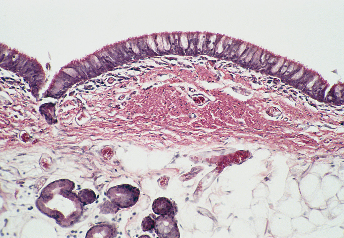
| Light micrograph of a longitudinal section of the trachea with lumen (white) at top. The epithelium (dark violet at top) is tall,pseudostratified and ciliated and contains numerous goblet cells (light violet). The epithelium is supported by a basement membrane which is not clearly visible here. Beneath this membrane there is a layer of loose,highly vascular connective tissue,the lamina propria,which becomes condensed at centre to form a band of fibro-elastic tissue. The loose submucosa (bottom) contains numerous mixed sero-mucous glands. A group of these is visible at bottom left. Magnification: x50 at 35mm size | |
| Licence : | Droits gérés |
| Crédit: | Science Photo Library / Michler, Astrid & Hans-Frieder |
| Taille de l’image : | 5195 px × 3592 px |
| Model Release : | Non requis |
| Property Release : | Non requis |
| Restrictions : | - |
Prix pour cette image À partir de 45 €
Produit vendu
(Calendrier, Carte postale, Carte de vœux, Impression sur textile, Packaging etc)
À partir de 45 €
Usage commercial
(Affichage, Annonce presse, Annonce TV, Carte, Digital - hors rés. sociaux, Digital - rés. sociaux etc)
À partir de 45 €
Éditorial
(Digital, Journal, Livre, Livre pratique, Magazine, Télévision etc)
À partir de 60 €
Usage non-commercial
(Digital - hors rés. sociaux, Digital - rés. sociaux etc)
À partir de 120 €