SEM of glandular surface of human colon
Numéro d’image : 11873283
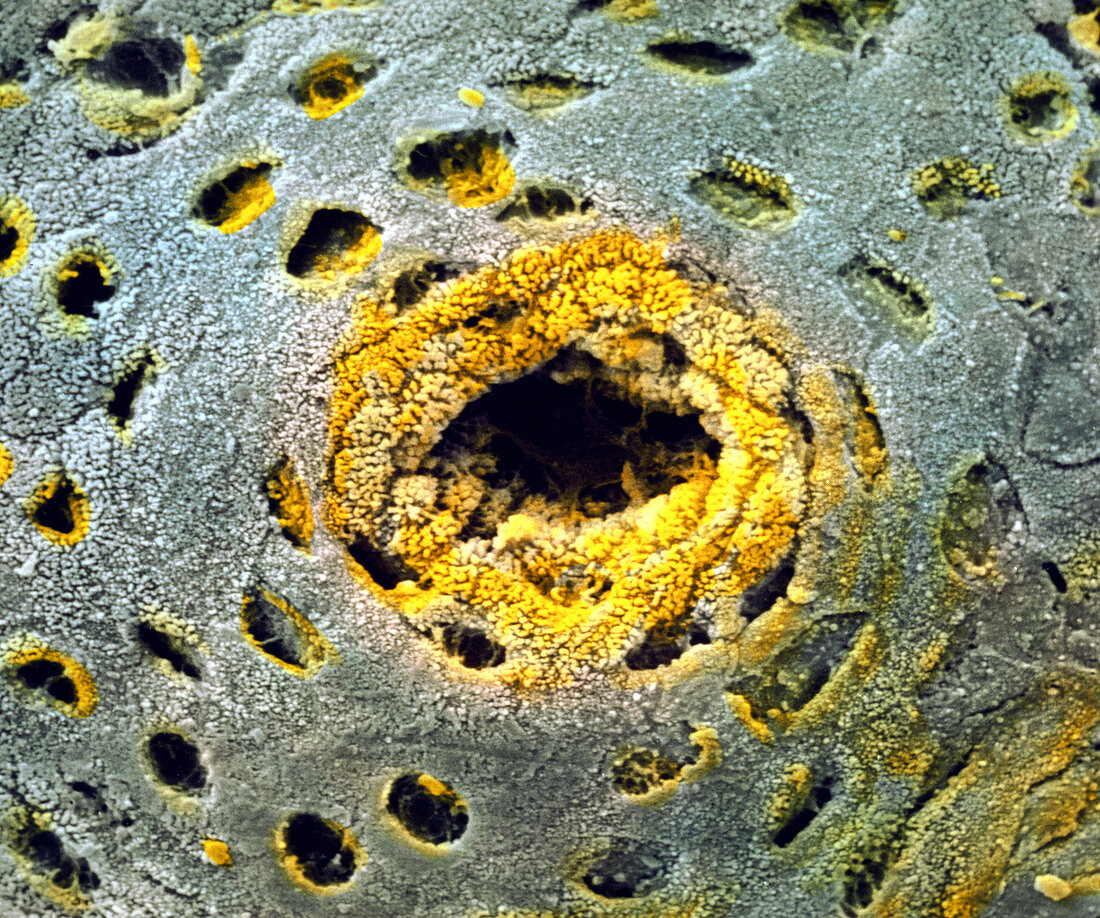
| Colon surface. Coloured Scanning Electron Micrograph (SEM) of the glandular surface of the wall (mucosa) of the human colon. At centre is a large opening or Crypt of Lieberkuhn of a tubular gland (yellow). Around it (blue) and making up the gland are two types of epithelial cells: enterocytes covered in tiny bumps (microvilli) that enhance water absorption; goblet cells that occur in the small rounded depressions seen here,and which secrete mucus. The glandular colon wall is lubricated with mucus,and functions to produce faeces by absorbing water and vitamins from the food residue. Magnification: x1,180 at 6x7cm size. x1,430 at 4x5ins | |
| Licence : | Droits gérés |
| Crédit: | Science Photo Library / PROFESSORS P.M. MOTTA & F.M. MAGLIOCCA |
| Taille de l’image : | 3543 px × 2957 px |
| Model Release : | Non requis |
| Property Release : | Non requis |
| Restrictions : | - |
Prix pour cette image À partir de 45 €
Produit vendu
(Calendrier, Carte postale, Carte de vœux, Impression sur textile, Packaging etc)
À partir de 45 €
Usage commercial
(Affichage, Annonce presse, Annonce TV, Carte, Digital - hors rés. sociaux, Digital - rés. sociaux etc)
À partir de 45 €
Éditorial
(Digital, Journal, Livre, Livre pratique, Magazine, Télévision etc)
À partir de 60 €
Usage non-commercial
(Digital - hors rés. sociaux, Digital - rés. sociaux etc)
À partir de 120 €