Gall bladder surface,SEM
Numéro d’image : 11872929
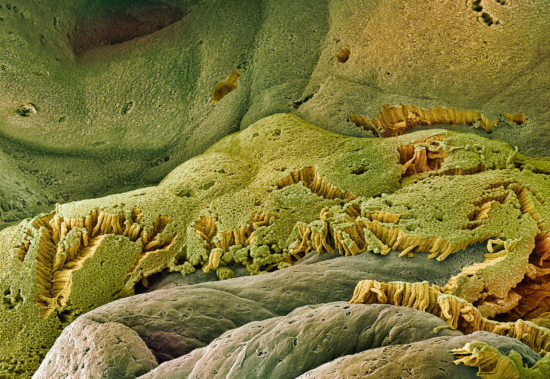
| Gall bladder. Coloured scanning electron micrograph (SEM) of the internal surface of a gall bladder. This mucosa lining is made up of columnar epithelial cells (green and yellow). Each cell of the gall bladder lining has microvilli (tiny projections) that increase its surface area and aid water uptake. Connective tissue (brown) is seen below the lining where some of the cells have come away. The gall bladder is a sac that concentrates and stores bile,produced by the liver,and releases it into the duodenum (small intestine),where it aids the digestion of fats. Magnification: x55 when printed 10 centimetres wide | |
| Licence : | Droits gérés |
| Crédit: | Science Photo Library / Gschmeissner, Steve |
| Taille de l’image : | 3500 px × 2411 px |
| Model Release : | Non requis |
| Property Release : | Non requis |
| Restrictions : | - |
Prix pour cette image À partir de 45 €
Produit vendu
(Calendrier, Carte postale, Carte de vœux, Impression sur textile, Packaging etc)
À partir de 45 €
Usage commercial
(Affichage, Annonce presse, Annonce TV, Carte, Digital - hors rés. sociaux, Digital - rés. sociaux etc)
À partir de 45 €
Éditorial
(Digital, Journal, Livre, Livre pratique, Magazine, Télévision etc)
À partir de 60 €
Usage non-commercial
(Digital - hors rés. sociaux, Digital - rés. sociaux etc)
À partir de 120 €
Mots clés
- anatomie,
- bile,
- biologie,
- biologique,
- cellule,
- cellule cylindrique,
- cellules,
- coloré,
- colorié,
- colorisé,
- corps humain,
- cylindrique,
- digestion,
- digestive,
- épithéliale,
- histologie,
- histologique,
- M.E.B.,
- MEB,
- microscope électronique à balayage,
- microvillosité,
- microvillus,
- mucosa,
- muqueuse,
- organe,
- stockage,
- surface,
- tissu conjonctif,
- vésicule biliaire