Coloured SEM of surface cells of the gall bladder
Numéro d’image : 11872890
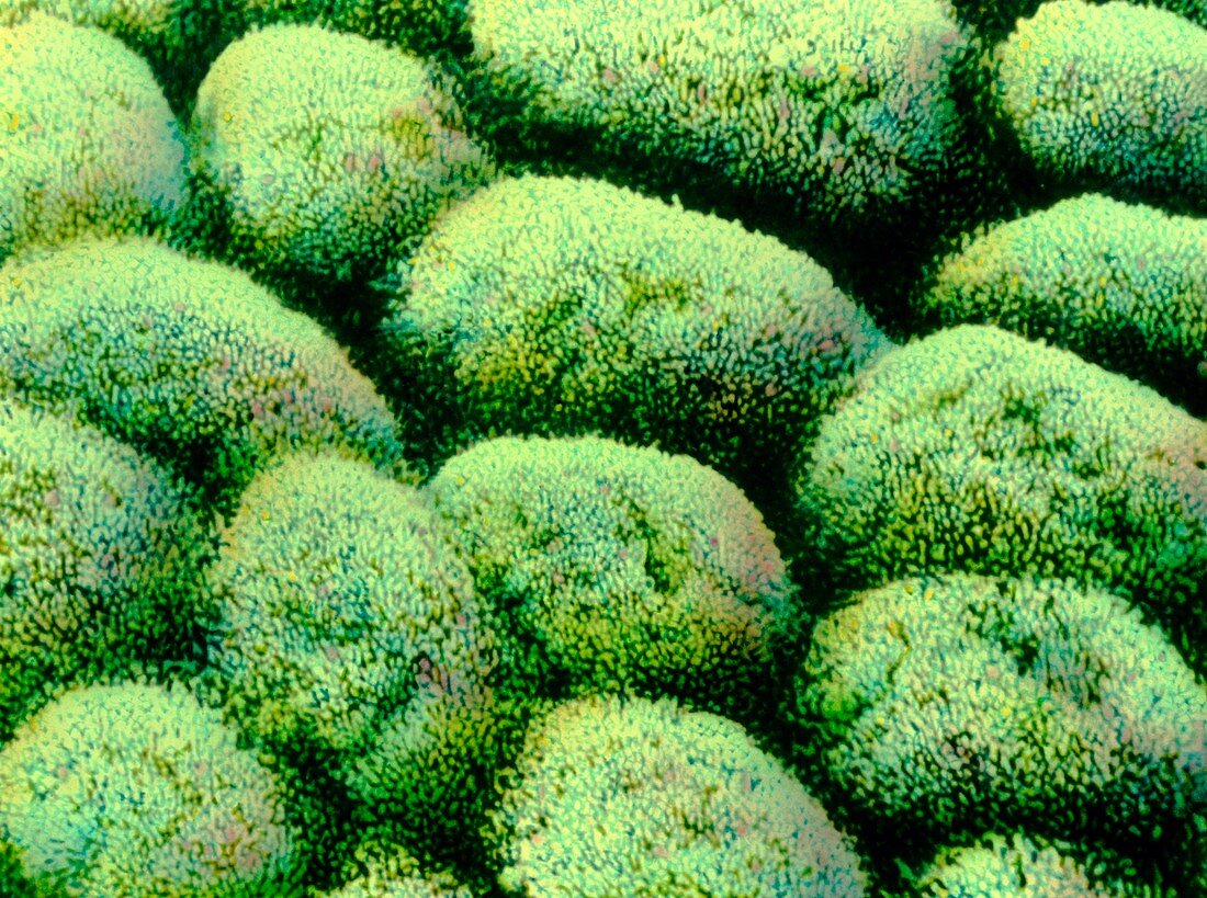
| Gall bladder. Coloured scanning electron micro- graph (SEM) of cells on the wall of the gall bladder. This lining mucosa is made up of columnar epithelial cells,each appearing rounded on the surface. Each cell is covered with small micro- villi (hair-like structures). Microvilli increase the surface area of these cells for absorbing water from bile. Beneath the folded mucosa wall lie sheets of smooth muscle. The gall bladder is a muscular sac attached to the liver. It collects & concentrates liver bile. Bile is then released from the gall bladder down the bile duct and into the duodenum of the intestine,where it aids in the digestion of food by emulsifying fats | |
| Licence : | Droits gérés |
| Crédit: | Science Photo Library / Gschmeissner, Steve |
| Taille de l’image : | 4837 px × 3602 px |
| Model Release : | Non requis |
| Property Release : | Non requis |
| Restrictions : | - |
Prix pour cette image À partir de 45 €
Produit vendu
(Calendrier, Carte postale, Carte de vœux, Impression sur textile, Packaging etc)
À partir de 45 €
Usage commercial
(Affichage, Annonce presse, Annonce TV, Carte, Digital - hors rés. sociaux, Digital - rés. sociaux etc)
À partir de 45 €
Éditorial
(Digital, Journal, Livre, Livre pratique, Magazine, Télévision etc)
À partir de 60 €
Usage non-commercial
(Digital - hors rés. sociaux, Digital - rés. sociaux etc)
À partir de 120 €