False-colour SEM of surface peritoneum of liver
Numéro d’image : 11872875
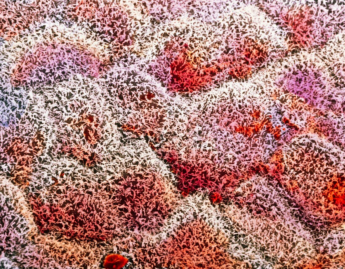
| False-colour scanning electron micrograph (SEM) of the surface peritoneal layer of cells around the liver. Covered with many short microvilli (hair- like structures),these flattened serosal cells form an organ holding sac around the liver. Its surface area,increased by the microvilli,secre- tes fluid derived from blood serum: this allows a frictionless movement of the liver within the abdomen. Such a protective layer for organs is important,especially in the abdomen,where many organs coexist and function during body movement. Beneath microvilli the outline of individual cells can be seen. Magnification: x1,125 at 6x7cm size. Magnification: x1,800 at 4x5 inch size | |
| Licence : | Droits gérés |
| Crédit: | Science Photo Library / UNIVERSITY LA SAPIENZA, ROME / DEPT. OF ANATOMY / PROF. P. MOTTA |
| Taille de l’image : | 4843 px × 3779 px |
| Model Release : | Non requis |
| Property Release : | Non requis |
| Restrictions : | - |
Prix pour cette image À partir de 45 €
Produit vendu
(Calendrier, Carte postale, Carte de vœux, Impression sur textile, Packaging etc)
À partir de 45 €
Usage commercial
(Affichage, Annonce presse, Annonce TV, Carte, Digital - hors rés. sociaux, Digital - rés. sociaux etc)
À partir de 45 €
Éditorial
(Digital, Journal, Livre, Livre pratique, Magazine, Télévision etc)
À partir de 60 €
Usage non-commercial
(Digital - hors rés. sociaux, Digital - rés. sociaux etc)
À partir de 120 €