False-colour SEM of cell structure of liver lobule
Numéro d’image : 11872864
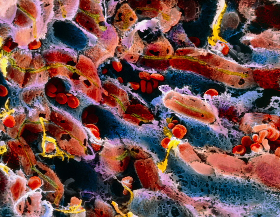
| False-colour scanning electron micrograph (SEM) of the cell structure within a lobule of the liver. Hepatic cells (brown) surround sinusoids (blue channels) in a three-dimensional network. Blood (red) flows through these sinusoids to link up to a central vein. In this process of blood flow,specialized liver cells clean the blood of microorganisms,toxins,aged cells,and debris. Bile formed by the liver cells is released into tiny bile capillaries (cholangioles,green). Bile enters the gall bladder to be used for digestive purposes. The liver also synthesizes Vitamin A. Magnification: x300 at 6x7cm size. Magnification: x460 at 4x5 inch size | |
| Licence : | Droits gérés |
| Crédit: | Science Photo Library / UNIVERSITY LA SAPIENZA, ROME / DEPT. OF ANATOMY / PROF. P. MOTTA |
| Taille de l’image : | 4975 px × 3864 px |
| Model Release : | Non requis |
| Property Release : | Non requis |
| Restrictions : | - |
Prix pour cette image À partir de 45 €
Produit vendu
(Calendrier, Carte postale, Carte de vœux, Impression sur textile, Packaging etc)
À partir de 45 €
Usage commercial
(Affichage, Annonce presse, Annonce TV, Carte, Digital - hors rés. sociaux, Digital - rés. sociaux etc)
À partir de 45 €
Éditorial
(Digital, Journal, Livre, Livre pratique, Magazine, Télévision etc)
À partir de 60 €
Usage non-commercial
(Digital - hors rés. sociaux, Digital - rés. sociaux etc)
À partir de 120 €