False-colour SEM of a lobule of the liver
Numéro d’image : 11872862
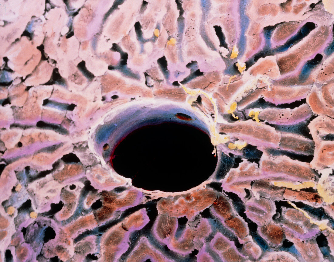
| False-colour scanning electron micrograph (SEM) of a lobule of the liver. Parenchyma tissue in the liver is arranged into lobules of cells,one of which is seen here. Each lobule has a central vein (black circle here); portal tracts at the boundary of each lobule (beyond this field of view) lead blood to flow along paths or sinusoids (grey) between hepatic cells,towards the centre of the lobule. In the process,blood is detoxified of waste products; proteins are metabolized; blood clotting agents,vitamin A and bile are synthe- sized; and spent red blood cells are reabsorbed. Magnification: x570 at 6x7cm size. Magnification: x875 at 4x5 inch size | |
| Licence : | Droits gérés |
| Crédit: | Science Photo Library / UNIVERSITY LA SAPIENZA, ROME / DEPT. OF ANATOMY / PROF. P. MOTTA |
| Taille de l’image : | 4345 px × 3401 px |
| Model Release : | Non requis |
| Property Release : | Non requis |
| Restrictions : | - |
Prix pour cette image À partir de 45 €
Produit vendu
(Calendrier, Carte postale, Carte de vœux, Impression sur textile, Packaging etc)
À partir de 45 €
Usage commercial
(Affichage, Annonce presse, Annonce TV, Carte, Digital - hors rés. sociaux, Digital - rés. sociaux etc)
À partir de 45 €
Éditorial
(Digital, Journal, Livre, Livre pratique, Magazine, Télévision etc)
À partir de 60 €
Usage non-commercial
(Digital - hors rés. sociaux, Digital - rés. sociaux etc)
À partir de 120 €