False-colour SEM of vessels in liver lobule
Numéro d’image : 11872858
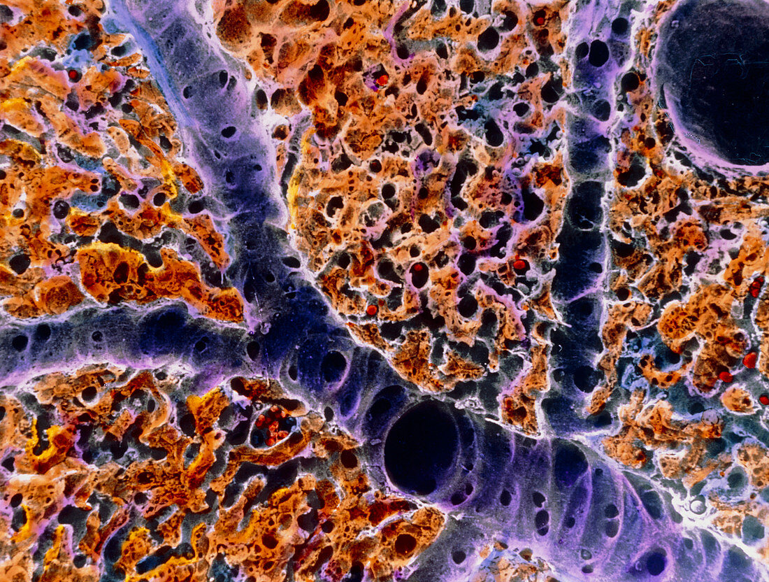
| Liver tissue. False-colour scanning electron micrograph (SEM) of blood vessels around a lobule,the functional unit of the liver. A typical lobule is bound on its periphery by groups of ducts & vessels,most prominently the conducting portal vein (large blue sectioned vessel) & branches called distributing portal veins (smaller blue vessel). Plates of hepatocytes or liver cells (coloured gold) radiate towards a central vein (top right). The dark spaces surrounding them are capillaries (sinusoids). Arterial vessels also bind each lobule,but are less visible here. Magnification: x125 at 6x7cm size | |
| Licence : | Droits gérés |
| Crédit: | Science Photo Library / UNIVERSITY LA SAPIENZA, ROME / DEPT. OF ANATOMY / PROF. P. MOTTA |
| Taille de l’image : | 3552 px × 2686 px |
| Model Release : | Non requis |
| Property Release : | Non requis |
| Restrictions : | - |
Prix pour cette image À partir de 45 €
Produit vendu
(Calendrier, Carte postale, Carte de vœux, Impression sur textile, Packaging etc)
À partir de 45 €
Usage commercial
(Affichage, Annonce presse, Annonce TV, Carte, Digital - hors rés. sociaux, Digital - rés. sociaux etc)
À partir de 45 €
Éditorial
(Digital, Journal, Livre, Livre pratique, Magazine, Télévision etc)
À partir de 60 €
Usage non-commercial
(Digital - hors rés. sociaux, Digital - rés. sociaux etc)
À partir de 120 €