Small intestine
Numéro d’image : 11872756
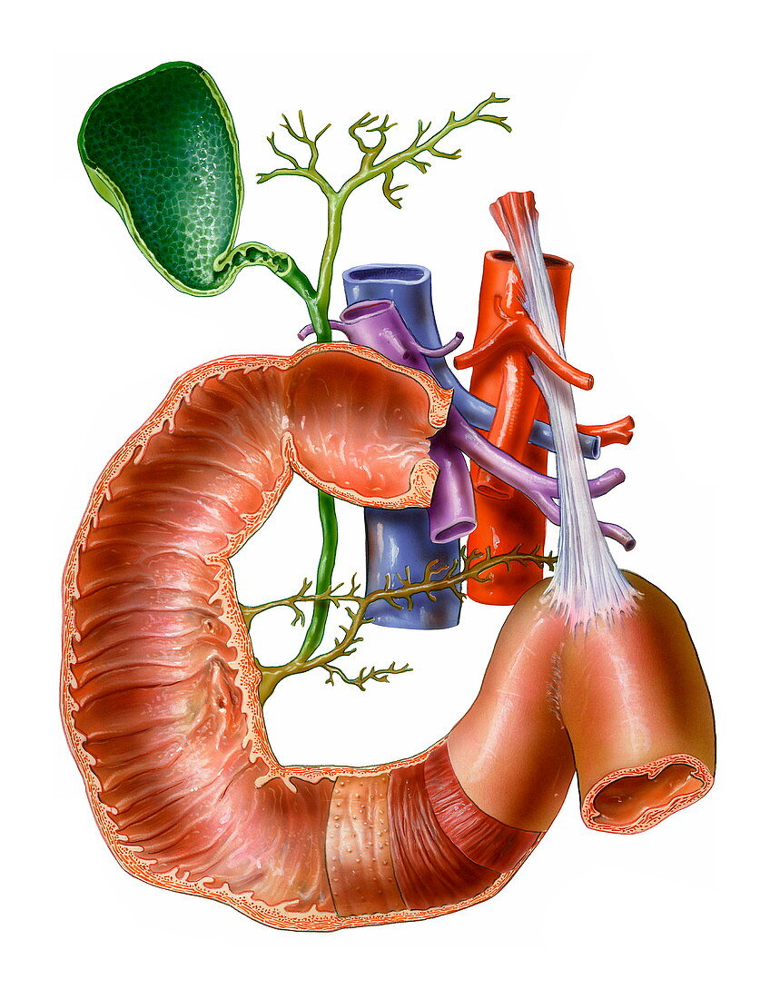
| Small intestine,artwork. At centre is the pylorus,the point where the stomach (not seen) opens into the duodenum,the first part of the small intestine. The duodenal wall contains plicae,small folds,which are covered in small projections called villi. These increase the surface area for digestion and absorption of food. At bottom centre are the layers of the duodenum wall,the submucosa,which contains blood vessels,circular muscles and longitudinal muscles. At centre is the portal vein (purple,which serves the liver),the vena cava (blue,the body's main vein) and the aorta (red,the body's main artery). The gall bladder (green) and pancreatic ducts (yellow) are also seen | |
| Licence : | Droits gérés |
| Crédit: | Science Photo Library / Veisland, Bo |
| Taille de l’image : | 2645 px × 3431 px |
| Model Release : | Non requis |
| Property Release : | Non requis |
| Restrictions : | - |
Prix pour cette image À partir de 45 €
Produit vendu
(Calendrier, Carte postale, Carte de vœux, Impression sur textile, Packaging etc)
À partir de 45 €
Usage commercial
(Affichage, Annonce presse, Annonce TV, Carte, Digital - hors rés. sociaux, Digital - rés. sociaux etc)
À partir de 45 €
Éditorial
(Digital, Journal, Livre, Livre pratique, Magazine, Télévision etc)
À partir de 60 €
Usage non-commercial
(Digital - hors rés. sociaux, Digital - rés. sociaux etc)
À partir de 120 €
Mots clés
- abdomen,
- anatomie,
- anatomique,
- aorta,
- aorte,
- appareil,
- artère,
- biologie,
- biologique,
- canal cholédoque,
- canaux pancréatiques,
- circulaire,
- conduits pancréatiques,
- corps humain,
- digestion,
- en bonne santé,
- illustration,
- intestin grêle,
- longitudinal,
- muscle,
- normal,
- oeuvre,
- PLICA,
- PLICAE,
- pylore,
- sain,
- système digestif,
- vaisseaux sanguins,
- veine,
- veine porte,
- veines,
- vésicule biliaire,
- voie,
- voie biliaire principale