Small intestine,TEM
Numéro d’image : 11872747
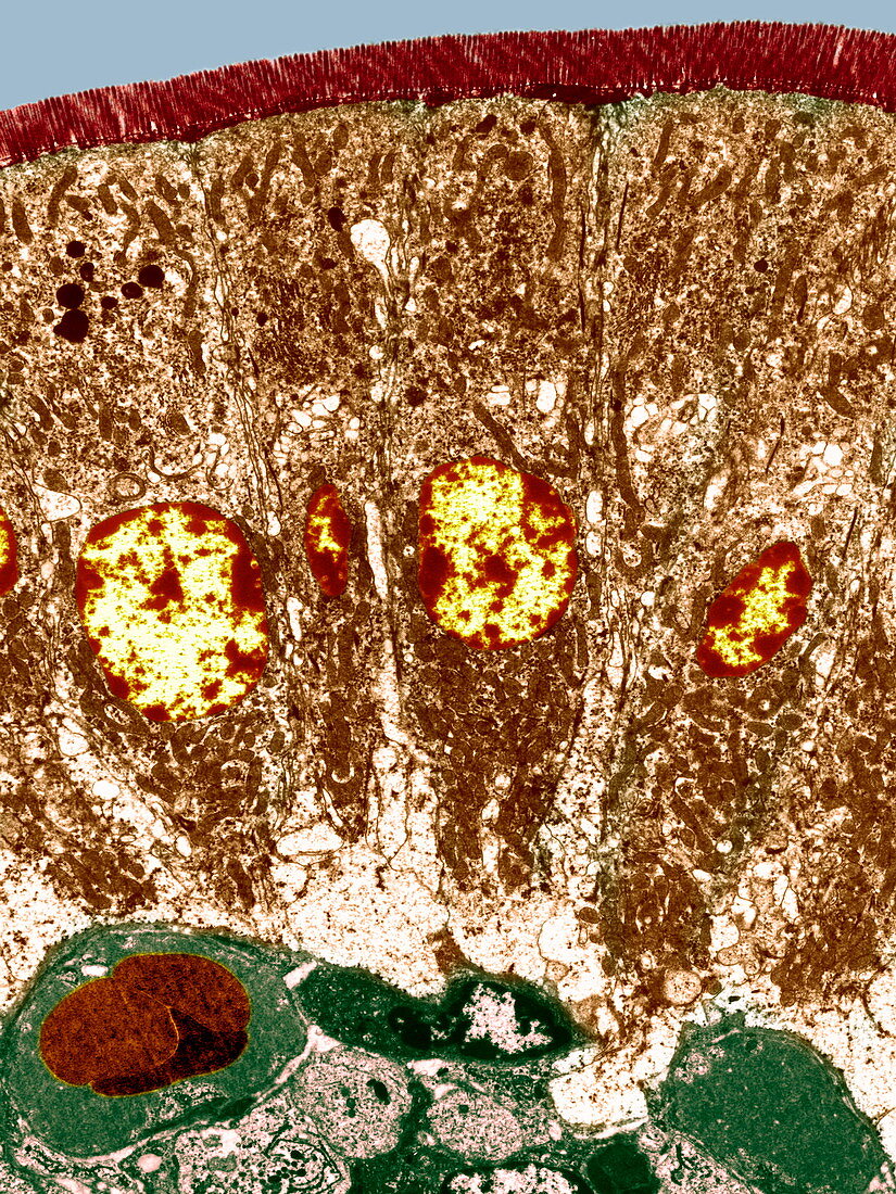
| Small intestine. Coloured transmission electron micrograph (TEM) of a section through the wall of the small intestine. The surface consists of many small hair-like absorptive structures (red) called microvilli. These absorb nutrients from food as it passes through the lumen (internal space,blue,across top). The microvilli are the upper layer of the surface (epithelial) cells. These are columnar in shape (and so are called columnar epithelial cells). These cells (three seen here) extend down to lower centre,and the nuclei (yellow) are seen. Magnification: x600 when printed 10cm high | |
| Licence : | Droits gérés |
| Crédit: | Science Photo Library / BIOMEDICAL IMAGING UNIT, SOUTHAMPTON GENERAL HOSPITAL |
| Taille de l’image : | 2700 px × 3600 px |
| Model Release : | Non requis |
| Property Release : | Non requis |
| Restrictions : | - |
Prix pour cette image À partir de 45 €
Produit vendu
(Calendrier, Carte postale, Carte de vœux, Impression sur textile, Packaging etc)
À partir de 45 €
Usage commercial
(Affichage, Annonce presse, Annonce TV, Carte, Digital - hors rés. sociaux, Digital - rés. sociaux etc)
À partir de 45 €
Éditorial
(Digital, Journal, Livre, Livre pratique, Magazine, Télévision etc)
À partir de 60 €
Usage non-commercial
(Digital - hors rés. sociaux, Digital - rés. sociaux etc)
À partir de 120 €
Mots clés
- anatomie,
- biologie,
- biologique,
- cellule,
- coloré,
- colorié,
- colorisé,
- digestion,
- digestive,
- duodénum,
- histologie,
- histologique,
- intestin grêle,
- intestinal,
- intestins,
- lubrification,
- M.E.T.,
- MET,
- micrographie,
- micrographie électronique à transmission,
- microscope,
- microscope électronique à transmission,
- physiologie,
- physiologique,
- tissus