Duodenum secretory cells
Numéro d’image : 11872700
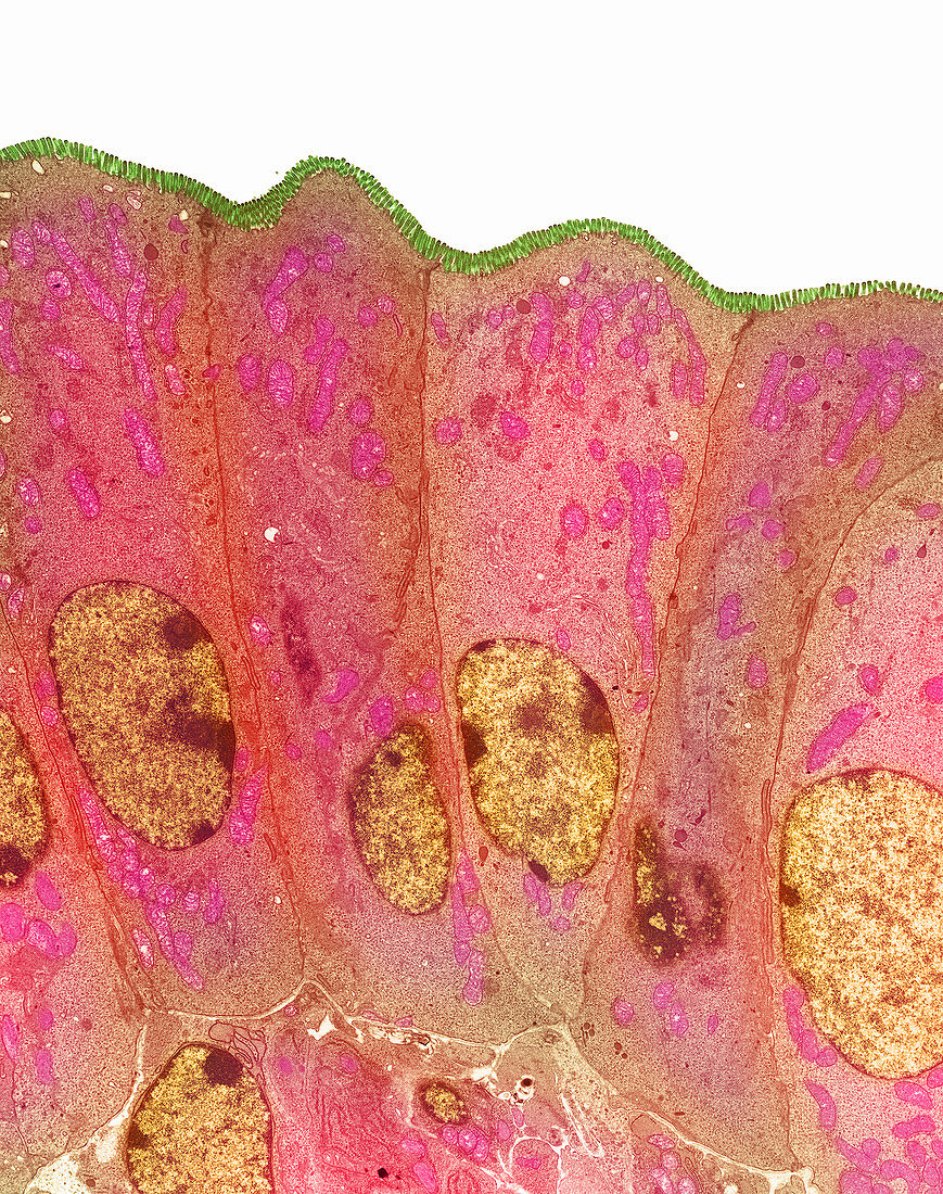
| Duodenum secretory cells. Coloured transmission electron micrograph (TEM) of a section through the human duodenum,showing secretory cells of the surface epithelium (lining). The duodenum is the first part of the small intestine. A row of columnar-shaped cells are seen,each with a rounded nucleus (brown) and mitochondria (pink) in the cytoplasm. Microvilli (green) appear as tiny projections from the surface of the cells (at top). Secretory cells secrete digestive enzymes,and an alkaline fluid into the pancreas which neutralises stomach acids. Microvilli serve to maximise the duodenum's surface area and hence its capacity to secrete. Magnification: unknown | |
| Licence : | Droits gérés |
| Crédit: | Science Photo Library / Gschmeissner, Steve |
| Taille de l’image : | 2765 px × 3500 px |
| Model Release : | Non requis |
| Property Release : | Non requis |
| Restrictions : | - |
Prix pour cette image À partir de 45 €
Produit vendu
(Calendrier, Carte postale, Carte de vœux, Impression sur textile, Packaging etc)
À partir de 45 €
Usage commercial
(Affichage, Annonce presse, Annonce TV, Carte, Digital - hors rés. sociaux, Digital - rés. sociaux etc)
À partir de 45 €
Éditorial
(Digital, Journal, Livre, Livre pratique, Magazine, Télévision etc)
À partir de 60 €
Usage non-commercial
(Digital - hors rés. sociaux, Digital - rés. sociaux etc)
À partir de 120 €