Artwork of a section through an intestinal villus
Numéro d’image : 11872684
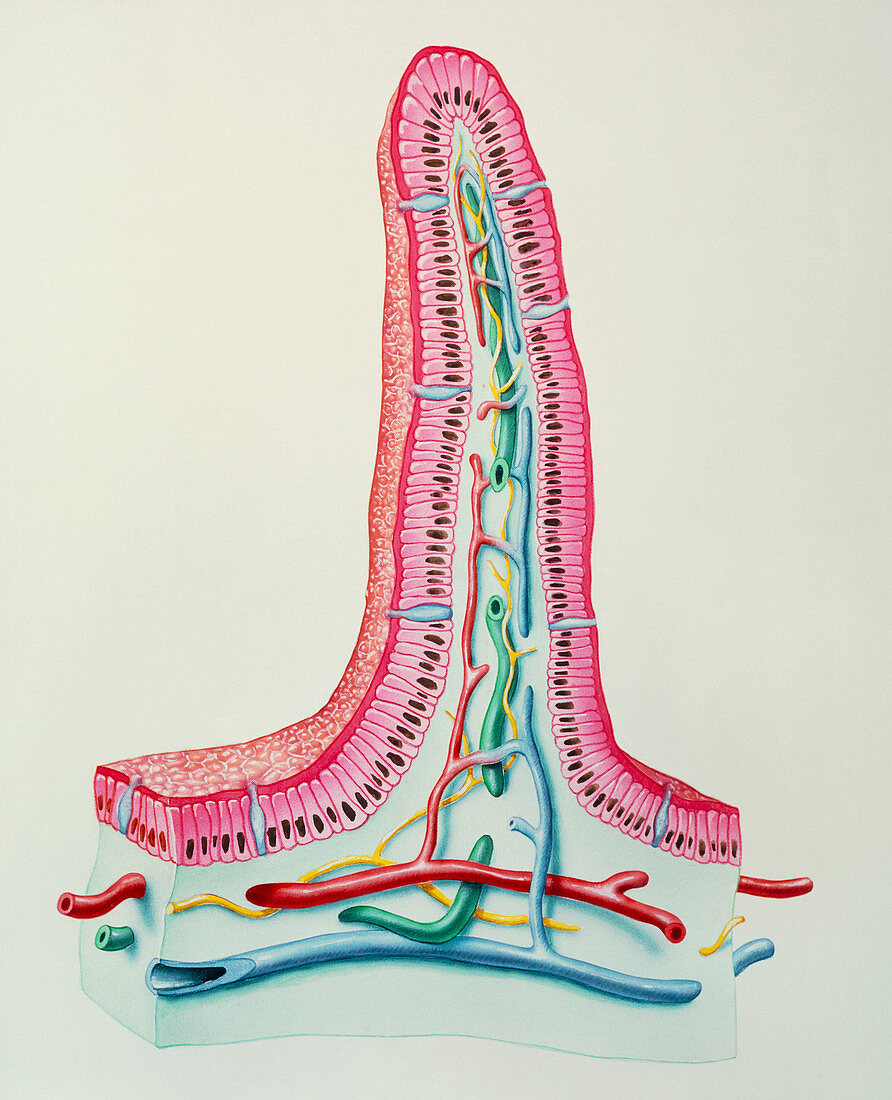
| Intestinal villus. Cutaway illustration of an intestinal villus. Villi are the finger-like projections that increase small intestine surface area. The villus is covered by columnar epithelium (pink). The epithelial cells are mostly enterocytes (with black nuclei) involved in digestion. Amongst the enterocytes are the goblet cells (blue) producing mucus which lubricates food and prevents self digestion. To the inside of the columnar epithelium is the lamina propria. This contains the arterial (red) & venous (blue) blood supply and lymph vessels (green). It is into the blood that the products of digestion are absorbed. Nerves (yellow) are also present | |
| Licence : | Droits gérés |
| Crédit: | Science Photo Library / BO VEISLAND, MI&I |
| Taille de l’image : | 4021 px × 4961 px |
| Model Release : | Non requis |
| Property Release : | Non requis |
| Restrictions : | - |
Prix pour cette image À partir de 45 €
Produit vendu
(Calendrier, Carte postale, Carte de vœux, Impression sur textile, Packaging etc)
À partir de 45 €
Usage commercial
(Affichage, Annonce presse, Annonce TV, Carte, Digital - hors rés. sociaux, Digital - rés. sociaux etc)
À partir de 45 €
Éditorial
(Digital, Journal, Livre, Livre pratique, Magazine, Télévision etc)
À partir de 60 €
Usage non-commercial
(Digital - hors rés. sociaux, Digital - rés. sociaux etc)
À partir de 120 €