LM of a section through the small intestine wall
Numéro d’image : 11872683
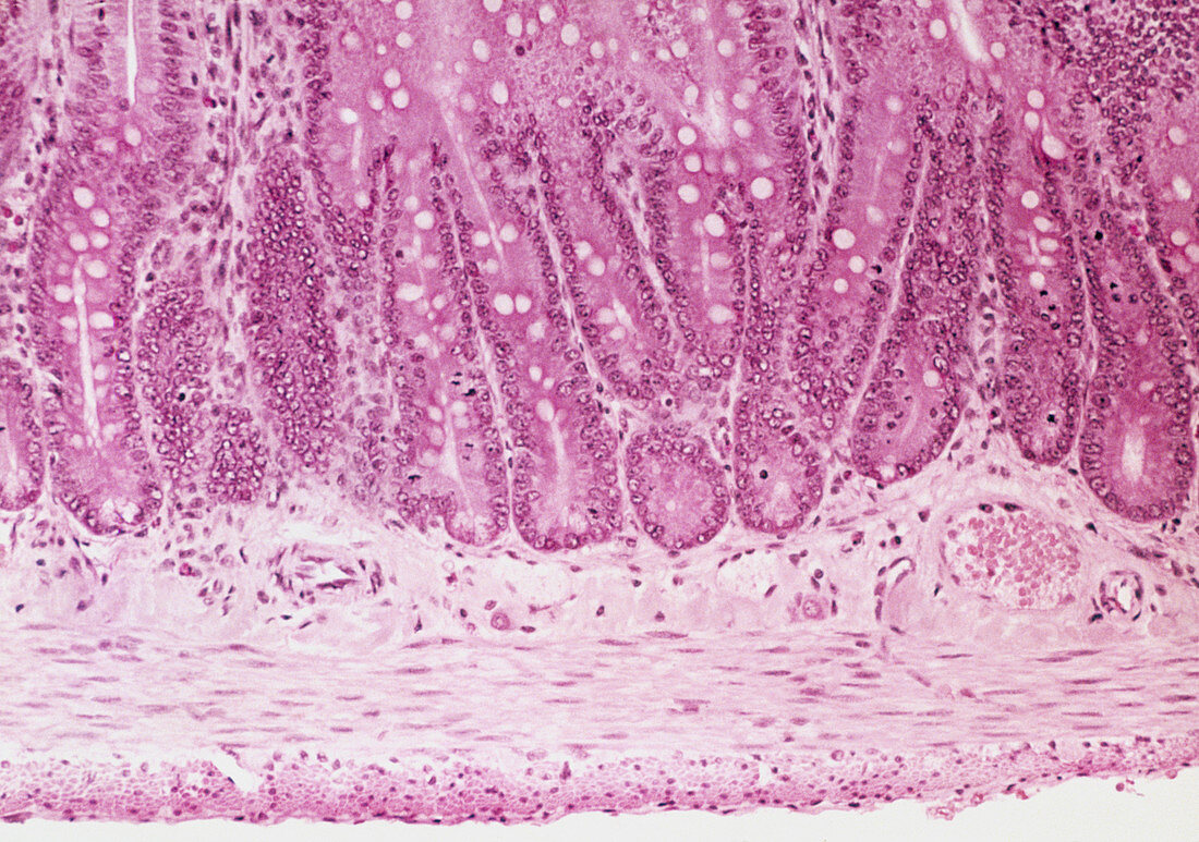
| Small intestine wall. Light micrograph of a section through the wall of the human small intestine. Making up the upper half of the frame are the crypts of Lieberkuhn (long,tubular) which lie at the base of the intestinal villi (unseen). They contain numerous mucus-producing goblet cells (white,round). Just below and between the crypts is the vascular submucosa. Beneath is the circular muscle layer lying above the longitudinal muscle layer. These layers of smooth muscle are responsible for the peristaltic action of the intestine. The thin outermost layer (at bottom) is the serosa composed of connective tissue. Magnification: x200 at 35mm size | |
| Licence : | Droits gérés |
| Crédit: | Science Photo Library / Science Pictures |
| Taille de l’image : | 5044 px × 3543 px |
| Model Release : | Non requis |
| Property Release : | Non requis |
| Restrictions : | - |
Prix pour cette image À partir de 45 €
Produit vendu
(Calendrier, Carte postale, Carte de vœux, Impression sur textile, Packaging etc)
À partir de 45 €
Usage commercial
(Affichage, Annonce presse, Annonce TV, Carte, Digital - hors rés. sociaux, Digital - rés. sociaux etc)
À partir de 45 €
Éditorial
(Digital, Journal, Livre, Livre pratique, Magazine, Télévision etc)
À partir de 60 €
Usage non-commercial
(Digital - hors rés. sociaux, Digital - rés. sociaux etc)
À partir de 120 €