Coloured TEM of microvilli of intestinal cell
Numéro d’image : 11872680
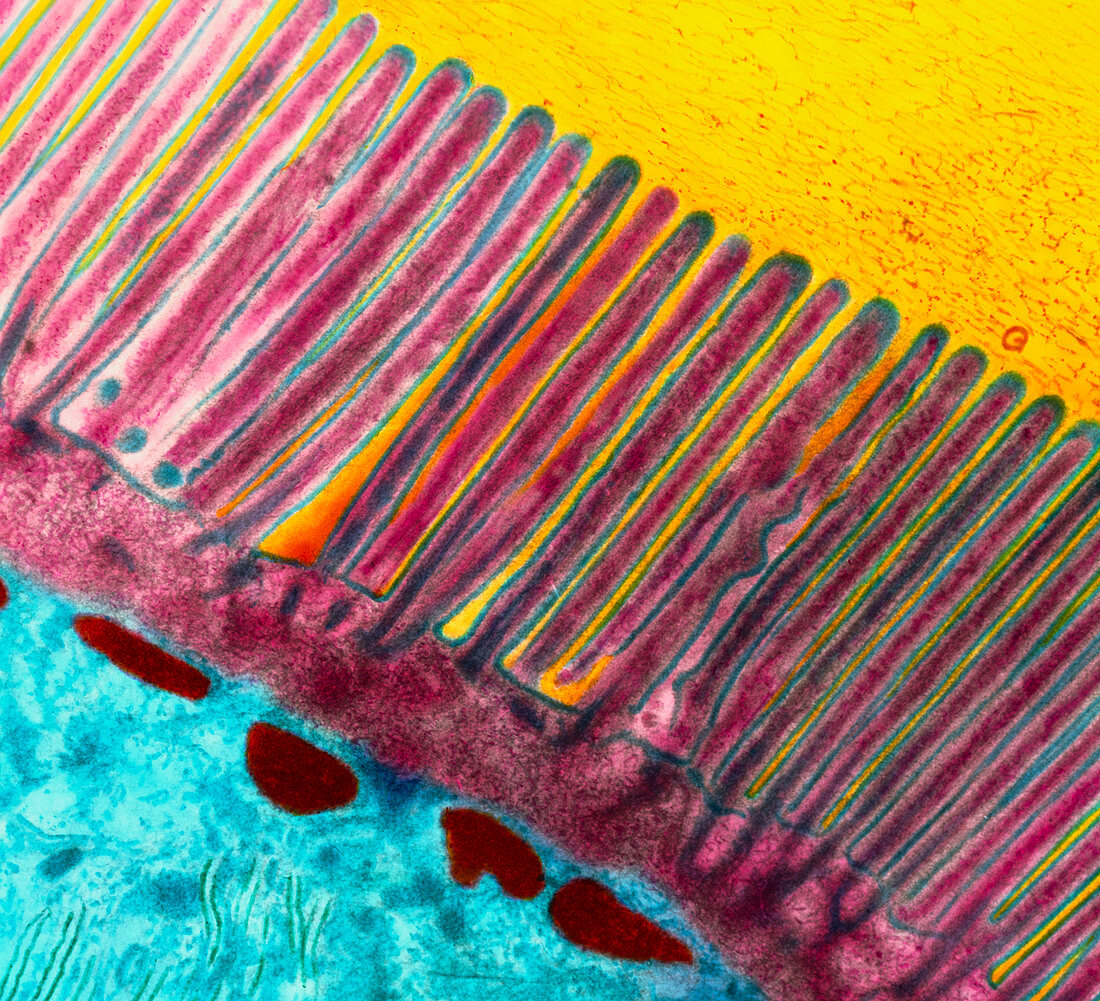
| Intestinal microvilli. Coloured transmission electron micrograph (TEM) of an epithelial cell from the small intestine showing microvilli. This intestinal cell absorbs nutrients from digested food. To increase the surface area available for absorption from the gut lumen (yellow) each cell is covered in thousands of microvilli (pink). These long finger-like projections together constitute a "brush border". The core (dark pink) of each microvillus is composed of microfilaments. These filaments contain contractile proteins providing primitive motility to the microvilli. Below them is the cell cytoplasm (blue). Magnification unknown | |
| Licence : | Droits gérés |
| Crédit: | Science Photo Library / Gschmeissner, Steve |
| Taille de l’image : | 4724 px × 4297 px |
| Model Release : | Non requis |
| Property Release : | Non requis |
| Restrictions : | - |
Prix pour cette image À partir de 45 €
Produit vendu
(Calendrier, Carte postale, Carte de vœux, Impression sur textile, Packaging etc)
À partir de 45 €
Usage commercial
(Affichage, Annonce presse, Annonce TV, Carte, Digital - hors rés. sociaux, Digital - rés. sociaux etc)
À partir de 45 €
Éditorial
(Digital, Journal, Livre, Livre pratique, Magazine, Télévision etc)
À partir de 60 €
Usage non-commercial
(Digital - hors rés. sociaux, Digital - rés. sociaux etc)
À partir de 120 €