LM of an intestinal villus in cross section
Numéro d’image : 11872675
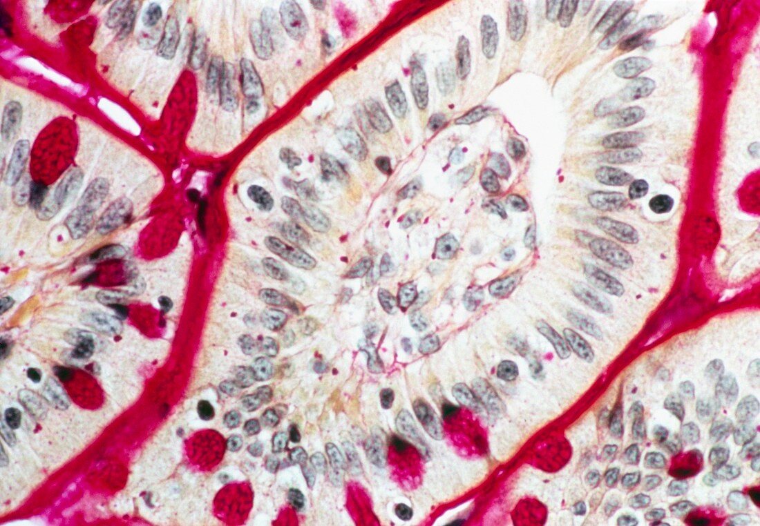
| Intestinal villus. Light micrograph of an intestinal villus in cross section. The villus is lined with simple columnar epithelium,containing two main types of cell. The enterocytes (pale brown with long grey nuclei) are involved in digestion and absorption. The goblet cells (pink) make and secrete mucus to lubricate food and protect against self digestion. In the centre of the villus is the lamina propria. This contains blood and lymph and is where digestive products are absorbed. Villi are folded projections of the lining of the small intestine. They greatly increase the surface area where absorption can take place. Stain and magnification unknown | |
| Licence : | Droits gérés |
| Crédit: | Science Photo Library / Biophoto Associates |
| Taille de l’image : | 5027 px × 3484 px |
| Model Release : | Non requis |
| Property Release : | Non requis |
| Restrictions : | - |
Prix pour cette image À partir de 45 €
Produit vendu
(Calendrier, Carte postale, Carte de vœux, Impression sur textile, Packaging etc)
À partir de 45 €
Usage commercial
(Affichage, Annonce presse, Annonce TV, Carte, Digital - hors rés. sociaux, Digital - rés. sociaux etc)
À partir de 45 €
Éditorial
(Digital, Journal, Livre, Livre pratique, Magazine, Télévision etc)
À partir de 60 €
Usage non-commercial
(Digital - hors rés. sociaux, Digital - rés. sociaux etc)
À partir de 120 €
Mots clés
- anatomie,
- cellule à mucus,
- cellule caliciforme,
- cellule cylindrique,
- cellule en gobelet,
- cellule mucipare,
- cellule muqueuse à pôle apical ouvert,
- cellules à mucus,
- corps humain,
- cylindrique,
- digestion,
- epithelium,
- épithélium,
- gut,
- intestin,
- intestin grêle,
- microscope optique,
- microscopie optique,
- petit,
- système digestif,
- villosité,
- villosités intestinales