Illustration of intestinal villi
Numéro d’image : 11872658
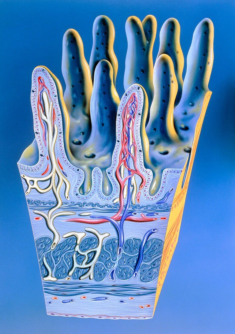
| Illustration of intestinal villi. These are minute finger-like projections found on the lining of the small intestine. Their role is food absorption. Each villus contains a lymph vessel (white) and a network of blood capillaries (red and blue). Tall columnar cells on the surface epithelium absorb food that passes over the villi. This food in turn passes into the blood circulation. Found at the base of villi are indentations called Crypts of Lieberkuhn. These crypts produce the cells that go to make up each villus. At bottom can be seen other muscle layers of the intestinal wall. Villi are largest and most numerous in the duodenum and jejunum,where most food absorption occurs | |
| Licence : | Droits gérés |
| Crédit: | Science Photo Library / Bavosi, John |
| Taille de l’image : | 3549 px × 5032 px |
| Model Release : | Non requis |
| Property Release : | Non requis |
| Restrictions : | - |
Prix pour cette image À partir de 45 €
Produit vendu
(Calendrier, Carte postale, Carte de vœux, Impression sur textile, Packaging etc)
À partir de 45 €
Usage commercial
(Affichage, Annonce presse, Annonce TV, Carte, Digital - hors rés. sociaux, Digital - rés. sociaux etc)
À partir de 45 €
Éditorial
(Digital, Journal, Livre, Livre pratique, Magazine, Télévision etc)
À partir de 60 €
Usage non-commercial
(Digital - hors rés. sociaux, Digital - rés. sociaux etc)
À partir de 120 €