SEM of cross-section through intestinal villi
Numéro d’image : 11872650
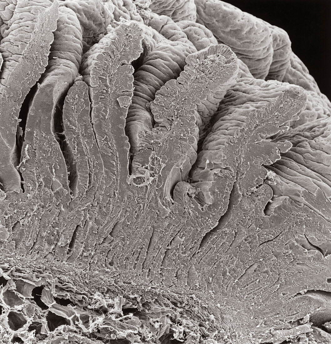
| Scanning electron micrograph of a section through the wall of the human duodenum,showing the many tiny folds,known as villi,which project 0.5 to 1 mm out into the intestinal lumen. These folds greatly increase the effective absorptive and secretory surface of the mucosa (mucus membrane) which lines the small intestine. Each villus contains a central core of connective tissue,known as the lamina propria (just visible as a strand of tissue running up the centre of the cut ends). This contains large blood vessels,capillaries,some smooth muscle cells and a blind- ended lymph vessel known as a lacteal. Magnification: x220 at 8x10 inch size | |
| Licence : | Droits gérés |
| Crédit: | Science Photo Library / Scharf, David |
| Taille de l’image : | 4134 px × 4295 px |
| Model Release : | Non requis |
| Property Release : | Non requis |
| Restrictions : |
|
Prix pour cette image À partir de 45 €
Produit vendu
(Calendrier, Carte postale, Carte de vœux, Impression sur textile, Packaging etc)
À partir de 45 €
Usage commercial
(Affichage, Annonce presse, Annonce TV, Carte, Digital - hors rés. sociaux, Digital - rés. sociaux etc)
À partir de 45 €
Éditorial
(Digital, Journal, Livre, Livre pratique, Magazine, Télévision etc)
À partir de 60 €
Usage non-commercial
(Digital - hors rés. sociaux, Digital - rés. sociaux etc)
À partir de 120 €