LM showing the wall of the human oesophagus
Numéro d’image : 11872560
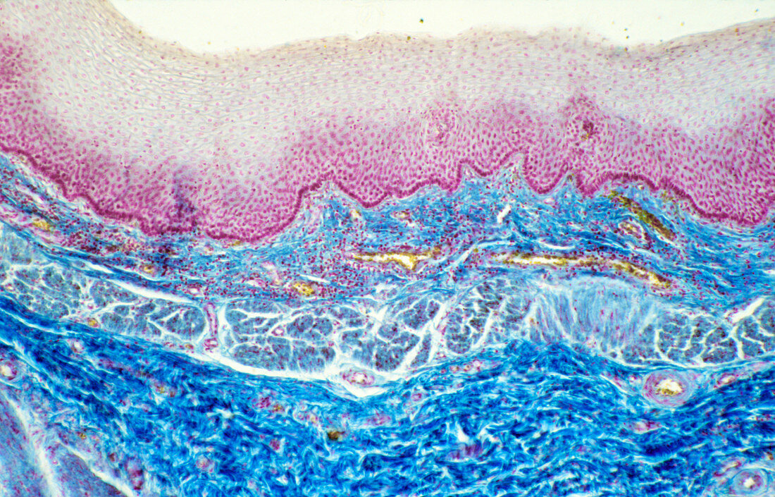
| Light micrograph of a vertical section through the wall of a human oesophagus. Its lumen is lined by a thick stratified squamous epithelium (grey and magenta). This is supported by a thin layer of connective tissue known as the lamina propria. At bottom the muscularis layer is visible. It consists of smooth muscle and is divided into two layers. The inner one (centre) is formed by circular fibres and the outer one (bottom,dark blue) by longitudinal fibres. The action of these two muscle layers,opposed at right angles to one another,is the basis of the peristaltic contraction. Magnification: x25 at 35mm size | |
| Licence : | Droits gérés |
| Crédit: | Science Photo Library / Michler, Astrid & Hans-Frieder |
| Taille de l’image : | 3895 px × 2496 px |
| Model Release : | Non requis |
| Property Release : | Non requis |
| Restrictions : | - |
Prix pour cette image À partir de 45 €
Produit vendu
(Calendrier, Carte postale, Carte de vœux, Impression sur textile, Packaging etc)
À partir de 45 €
Usage commercial
(Affichage, Annonce presse, Annonce TV, Carte, Digital - hors rés. sociaux, Digital - rés. sociaux etc)
À partir de 45 €
Éditorial
(Digital, Journal, Livre, Livre pratique, Magazine, Télévision etc)
À partir de 60 €
Usage non-commercial
(Digital - hors rés. sociaux, Digital - rés. sociaux etc)
À partir de 120 €