False-colour SEM of epithelium in the oesophagus
Numéro d’image : 11872558
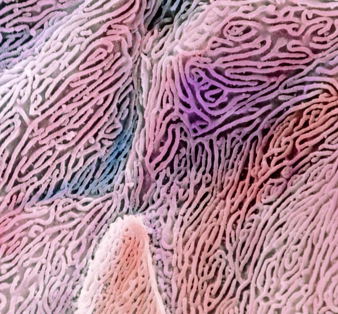
| False-colour scanning electron micrograph (SEM) of the epithelium in the oesophagus. Individual epithelial cells are seen here,each with a highly folded surface. These microfolds are called micro- plicae. At lower centre left,an obvious boundary to one cell can be seen. This stratified squamous epithelium consists of flattened cells that occur many layers thick. As a muscular non-absorptive tube,the oesophagus transports swallowed food to the stomach. The microfolds on the cells keep the oesophagus moist; trap mucous to lubricate passing food; and strengthen the epithelium against food abrasion. Magnification: x2,900 at 6x7cm size. Magnification: x4,650 at 4x5 inch size | |
| Licence : | Droits gérés |
| Crédit: | Science Photo Library / UNIVERSITY LA SAPIENZA, ROME / DEPT. OF ANATOMY / PROF. P. MOTTA |
| Taille de l’image : | 4382 px × 4073 px |
| Model Release : | Non requis |
| Property Release : | Non requis |
| Restrictions : | - |
Prix pour cette image À partir de 45 €
Produit vendu
(Calendrier, Carte postale, Carte de vœux, Impression sur textile, Packaging etc)
À partir de 45 €
Usage commercial
(Affichage, Annonce presse, Annonce TV, Carte, Digital - hors rés. sociaux, Digital - rés. sociaux etc)
À partir de 45 €
Éditorial
(Digital, Journal, Livre, Livre pratique, Magazine, Télévision etc)
À partir de 60 €
Usage non-commercial
(Digital - hors rés. sociaux, Digital - rés. sociaux etc)
À partir de 120 €