Larynx,3-D CT scan
Numéro d’image : 11872492
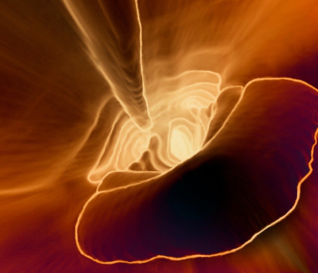
| Larynx. Coloured three-dimensional (3-D) computed tomography (CT) scan showing the epiglottis (lower right) and the descending larynx. The epiglottis is a flap of tissue that separates the trachea (windpipe) from the oesophagus (gullet). It flips between two positions to prevent food from entering the lungs,and air from entering the stomach. The larynx is the part of the windpipe that houses the vocal cords (not seen),which vibrate and tighten to help to modify sounds. CT scanning uses computers to build up 3-D images from a series of X-ray images | |
| Licence : | Droits gérés |
| Crédit: | Science Photo Library / Zephyr |
| Taille de l’image : | 3969 px × 3402 px |
| Model Release : | Non requis |
| Property Release : | Non requis |
| Restrictions : | - |
Prix pour cette image À partir de 45 €
Produit vendu
(Calendrier, Carte postale, Carte de vœux, Impression sur textile, Packaging etc)
À partir de 45 €
Usage commercial
(Affichage, Annonce presse, Annonce TV, Carte, Digital - hors rés. sociaux, Digital - rés. sociaux etc)
À partir de 45 €
Éditorial
(Digital, Journal, Livre, Livre pratique, Magazine, Télévision etc)
À partir de 60 €
Usage non-commercial
(Digital - hors rés. sociaux, Digital - rés. sociaux etc)
À partir de 120 €