Light micrograph of the parotid gland
Numéro d’image : 11872377
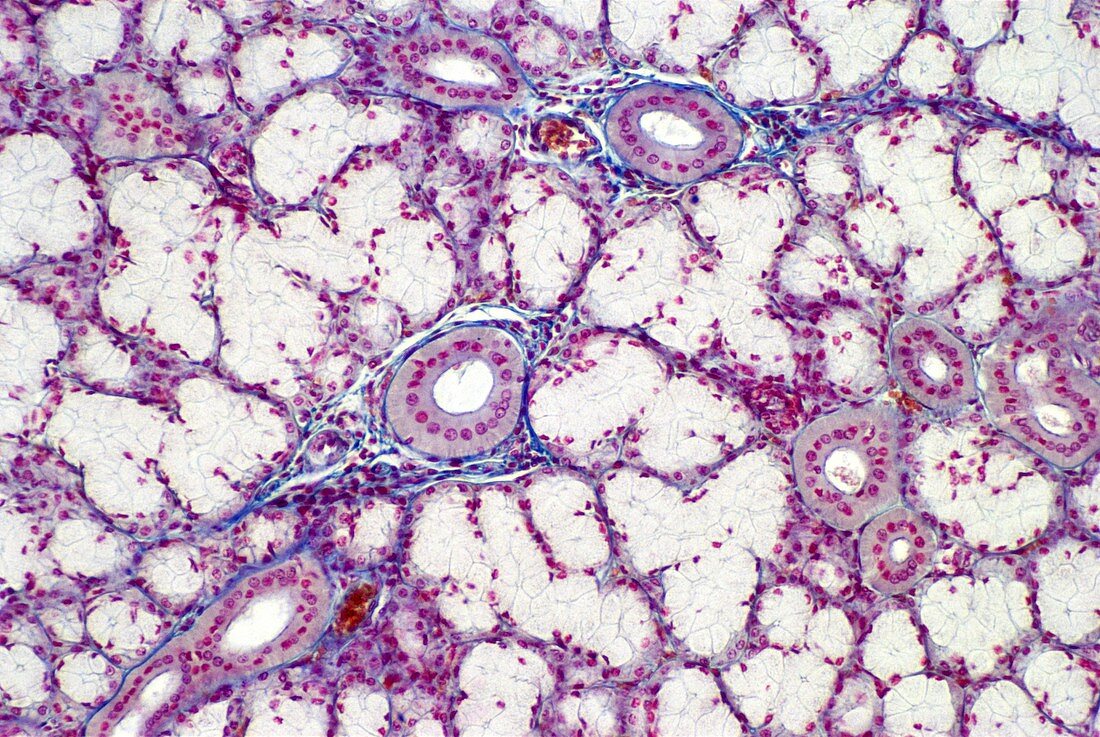
| Light micrograph of normal human parotid gland. One of the salivary glands,the parotid consists of acini whose secretion is entirely serous (watery). Most of the field here shows,in cross section,the secretory cells which comprise the acini. The prominent ring-shaped structures with many red nuclei and faint radial striping at their periphery are striated ducts. The blue stained cells forming boundaries around the striated ducts are conncetive tissue. Three blood vessels are visible,lying next to the ducts (e.g. centre top),containing orange stained red blood cells. Magnification: x50 at 35mm size | |
| Licence : | Droits gérés |
| Crédit: | Science Photo Library / Michler, Astrid & Hans-Frieder |
| Taille de l’image : | 5152 px × 3454 px |
| Model Release : | Non requis |
| Property Release : | Non requis |
| Restrictions : | - |
Prix pour cette image À partir de 45 €
Produit vendu
(Calendrier, Carte postale, Carte de vœux, Impression sur textile, Packaging etc)
À partir de 45 €
Usage commercial
(Affichage, Annonce presse, Annonce TV, Carte, Digital - hors rés. sociaux, Digital - rés. sociaux etc)
À partir de 45 €
Éditorial
(Digital, Journal, Livre, Livre pratique, Magazine, Télévision etc)
À partir de 60 €
Usage non-commercial
(Digital - hors rés. sociaux, Digital - rés. sociaux etc)
À partir de 120 €