Coloured SEM of filiform papillae on the tongue
Numéro d’image : 11872343
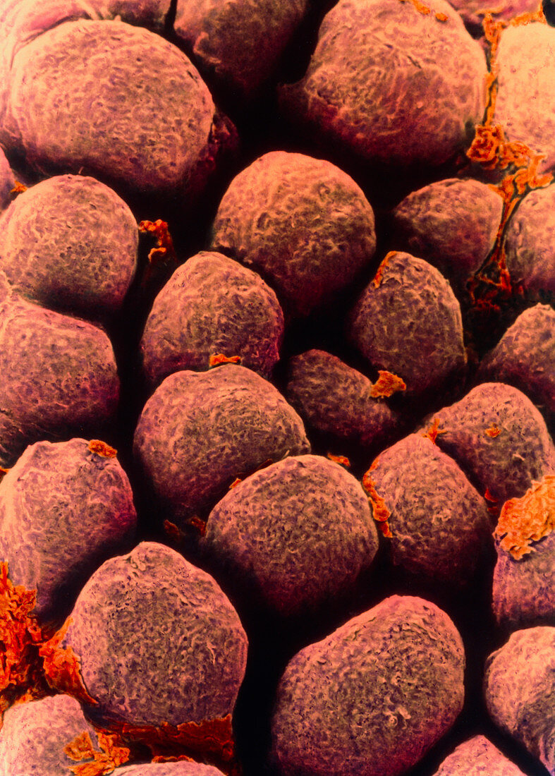
| Tongue papillae. Coloured scanning electron micrograph (SEM) of filiform papillae on the surface of the tongue. Filiform papillae are covered by stratified squamous epithelial cells. Dead cells of the uppermost layer are constantly being shed and replaced (desquamation). This shedding gives the papillae their scaly appearance. Filiform papillae form a rough surface to aid chewing. Each papilla contains nerve endings which transmit tactile (touch) information to the brain. Magnification: x57 at 5x7cm size. x200 at 7x9.5ins | |
| Licence : | Droits gérés |
| Crédit: | Science Photo Library / Gschmeissner, Steve |
| Taille de l’image : | 3572 px × 4993 px |
| Model Release : | Non requis |
| Property Release : | Non requis |
| Restrictions : | - |
Prix pour cette image À partir de 45 €
Produit vendu
(Calendrier, Carte postale, Carte de vœux, Impression sur textile, Packaging etc)
À partir de 45 €
Usage commercial
(Affichage, Annonce presse, Annonce TV, Carte, Digital - hors rés. sociaux, Digital - rés. sociaux etc)
À partir de 45 €
Éditorial
(Digital, Journal, Livre, Livre pratique, Magazine, Télévision etc)
À partir de 60 €
Usage non-commercial
(Digital - hors rés. sociaux, Digital - rés. sociaux etc)
À partir de 120 €