False-colour SEM of filiform papilla with bacteria
Numéro d’image : 11872327
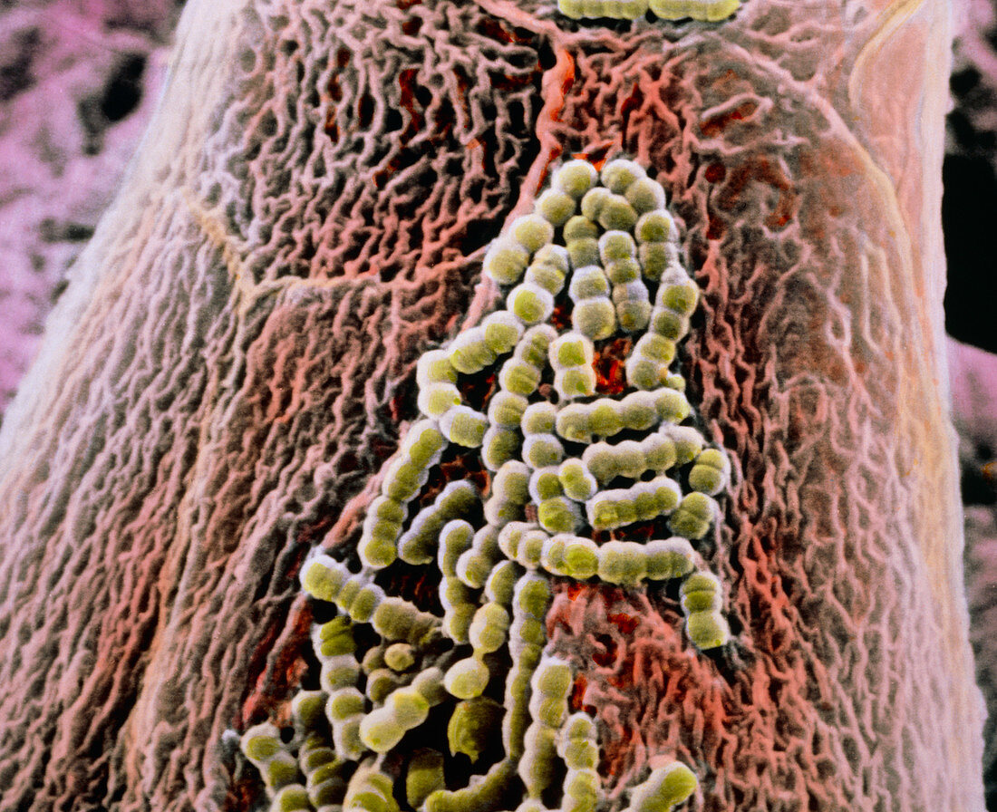
| False-colour scanning electron micrograph of part of a filiform papilla emerging from the tongue's surface. It is partly covered by green coloured bacteria. Filiform papillae,also known as conical papillae,are covered by stratified squamous epithelial cells apart from their tips,which are formed by the fibrous protein keratin in order to increase the papillae's strength. The image clearly shows the intricate pattern of microfolds on the cell surfaces. Filiform papillae have mechanical and tactile functions and form a rough surface which helps mastication. Magnification: x5290 at 6x7cm size. Magnification: x8050 at 4x5 inch size | |
| Licence : | Droits gérés |
| Crédit: | Science Photo Library / UNIVERSITY LA SAPIENZA, ROME / DEPT. OF ANATOMY / PROF. P. MOTTA |
| Taille de l’image : | 3457 px × 2816 px |
| Model Release : | Non requis |
| Property Release : | Non requis |
| Restrictions : | - |
Prix pour cette image À partir de 45 €
Produit vendu
(Calendrier, Carte postale, Carte de vœux, Impression sur textile, Packaging etc)
À partir de 45 €
Usage commercial
(Affichage, Annonce presse, Annonce TV, Carte, Digital - hors rés. sociaux, Digital - rés. sociaux etc)
À partir de 45 €
Éditorial
(Digital, Journal, Livre, Livre pratique, Magazine, Télévision etc)
À partir de 60 €
Usage non-commercial
(Digital - hors rés. sociaux, Digital - rés. sociaux etc)
À partir de 120 €