False-colour SEM of fungiform papillae on tongue
Numéro d’image : 11872324
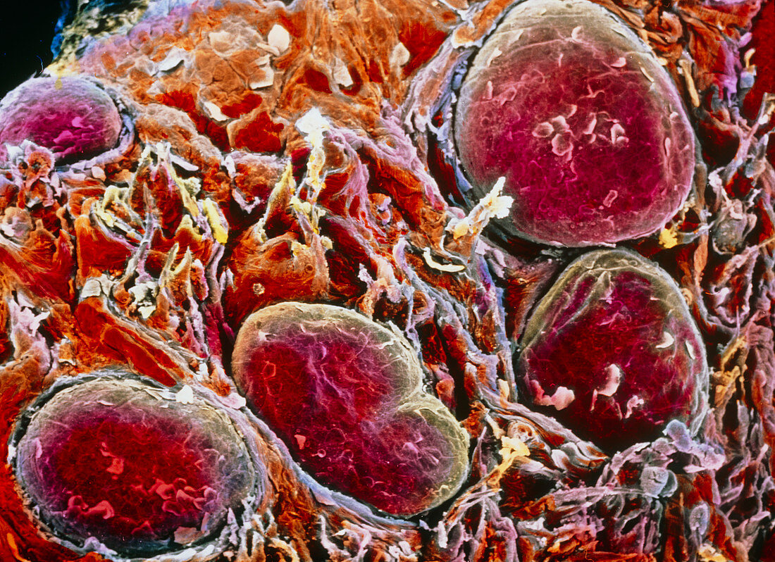
| Tongue surface. False-colour scanning electron micrograph of fungiform papillae (round & red) interspersed with a few conical-shaped filiform papillae. Fungiform papillae appear pinker because of the numerous blood vessels in the underlying connective tissue. Fungiform & filiform papillae are covered by layers of stratified squamous epithelium. Dead cells are constantly shed & replaced by cells of the underlying layers. Taste buds (not visible here) are usually found under the surface of fungiform papillae. Filiform papillae have tactile & mechanical functions. Magnification: x95 at 6x7cm size. Magnification: x155 at 4x5 inch size | |
| Licence : | Droits gérés |
| Crédit: | Science Photo Library / UNIVERSITY LA SAPIENZA, ROME / DEPT. OF ANATOMY / PROF. P. MOTTA |
| Taille de l’image : | 5070 px × 3678 px |
| Model Release : | Non requis |
| Property Release : | Non requis |
| Restrictions : | - |
Prix pour cette image À partir de 45 €
Produit vendu
(Calendrier, Carte postale, Carte de vœux, Impression sur textile, Packaging etc)
À partir de 45 €
Usage commercial
(Affichage, Annonce presse, Annonce TV, Carte, Digital - hors rés. sociaux, Digital - rés. sociaux etc)
À partir de 45 €
Éditorial
(Digital, Journal, Livre, Livre pratique, Magazine, Télévision etc)
À partir de 60 €
Usage non-commercial
(Digital - hors rés. sociaux, Digital - rés. sociaux etc)
À partir de 120 €