False colour SEM of a filiform papilla on tongue
Numéro d’image : 11872318
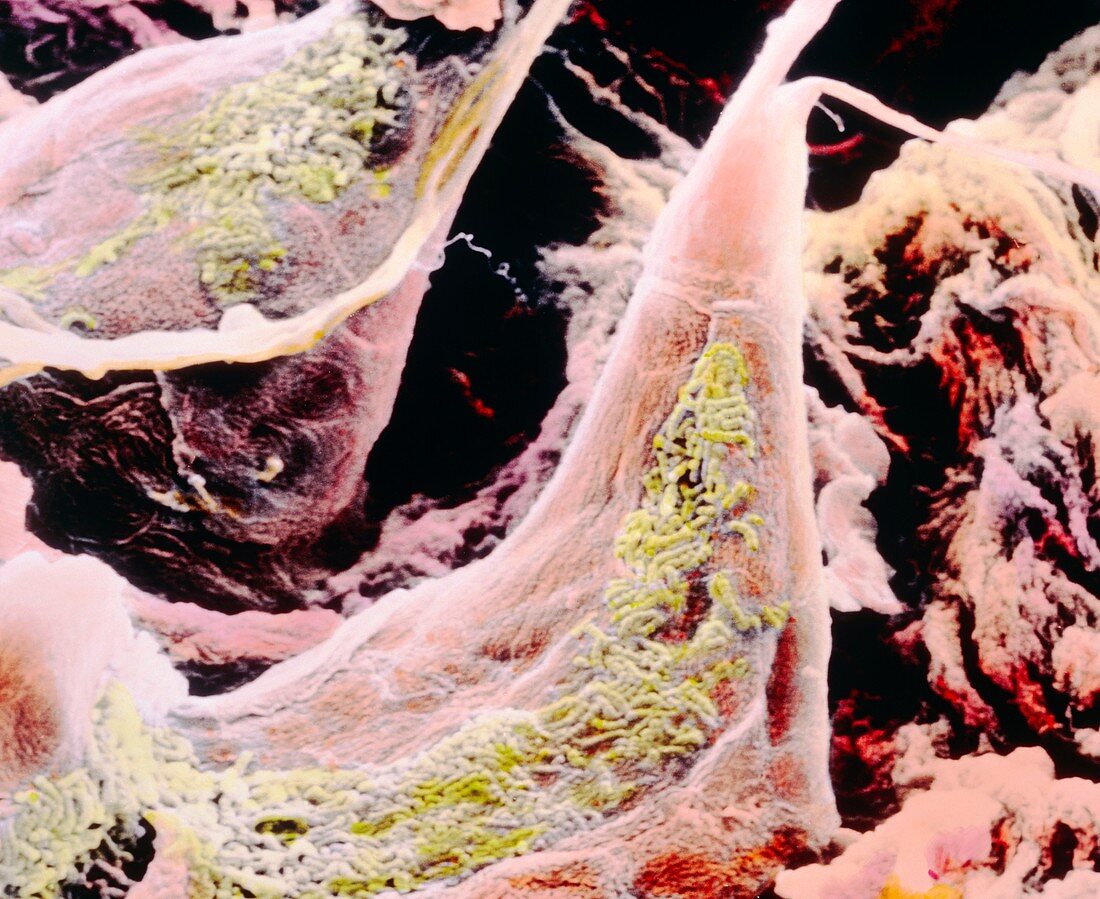
| False-colour scanning electron micrograph (SEM) of a filiform papilla emerging from the tongue's surface. It is partly covered by green coloured bacteria which may derive from a mouth infection. Filiform papillae,also known as conical papillae,are covered by epithelial cells apart from their white tips which are formed by the fibrous protein keratin in order to increase the papillae's strength. Filiform papillae have mechanical and tactile functions. They form a rough surface which helps mastication. Each papilla contains nerve endings which transmit tactile information to the brain. Magnification: x1100 at 6x7cm size | |
| Licence : | Droits gérés |
| Crédit: | Science Photo Library / UNIVERSITY LA SAPIENZA, ROME / DEPT. OF ANATOMY / PROF. P. MOTTA |
| Taille de l’image : | 4683 px × 3827 px |
| Model Release : | Non requis |
| Property Release : | Non requis |
| Restrictions : | - |
Prix pour cette image À partir de 45 €
Produit vendu
(Calendrier, Carte postale, Carte de vœux, Impression sur textile, Packaging etc)
À partir de 45 €
Usage commercial
(Affichage, Annonce presse, Annonce TV, Carte, Digital - hors rés. sociaux, Digital - rés. sociaux etc)
À partir de 45 €
Éditorial
(Digital, Journal, Livre, Livre pratique, Magazine, Télévision etc)
À partir de 60 €
Usage non-commercial
(Digital - hors rés. sociaux, Digital - rés. sociaux etc)
À partir de 120 €