Foetal inner ear hair cells,SEM
Numéro d’image : 11872162
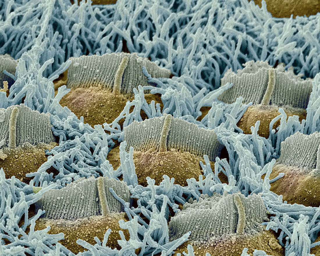
| Foetal inner ear hair cells. Coloured scanning electron micrograph (SEM) of hair cells in the organ of Corti of a foetus. This delicate structure in the cochlea of the inner ear converts sound vibrations into nerve impulses. The three rows of outer hair cells have V-shaped groups of stereocilia,each with a central kinocilium. In the foetus they are surrounded by numerous microvilli (blue),which are resorbed in the adult. The hair cells are surrounded by endolymph fluid. As sound enters the ear it causes waves to form in the endolymph,which in turn cause these hairs to move. The movement is converted into nerve impulses that are passed to the brain | |
| Licence : | Droits gérés |
| Crédit: | Science Photo Library / Gschmeissner, Steve |
| Taille de l’image : | 3500 px × 2800 px |
| Model Release : | Non requis |
| Property Release : | Non requis |
| Restrictions : | - |
Prix pour cette image À partir de 45 €
Produit vendu
(Calendrier, Carte postale, Carte de vœux, Impression sur textile, Packaging etc)
À partir de 45 €
Usage commercial
(Affichage, Annonce presse, Annonce TV, Carte, Digital - hors rés. sociaux, Digital - rés. sociaux etc)
À partir de 45 €
Éditorial
(Digital, Journal, Livre, Livre pratique, Magazine, Télévision etc)
À partir de 60 €
Usage non-commercial
(Digital - hors rés. sociaux, Digital - rés. sociaux etc)
À partir de 120 €
Mots clés
- anatomie,
- audition,
- cellule ciliée,
- cellules,
- cil,
- cils,
- cochlea,
- cochléaire,
- cochlée,
- colonne,
- coloré,
- colorié,
- colorisé,
- corps humain,
- extérieur,
- externe,
- foetal,
- foetale,
- foetus,
- intérieur,
- interne,
- intime,
- kinocilia,
- kinocilium,
- M.E.B.,
- MEB,
- microscope électronique à balayage,
- microvillosité,
- microvillus,
- oreille,
- organe de Corti,
- pilier,
- sens auditif,
- sensible,
- sensoriel,
- STEREOCILIA