Cochlea cells,SEM
Numéro d’image : 11872148
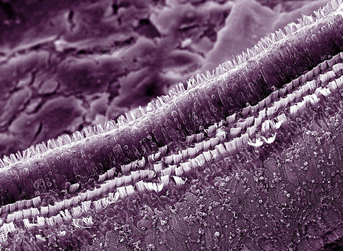
| Cochlea cells. Scanning electron micrograph (SEM) of a section through part of the cochlea inside a human ear. The rows of columnar outer pillar cells run along the organ of Corti,the auditory sense organ. The outer pillar cells arise from the basilar membrane,and their upper surfaces form part of the surface of the organ of Corti. This organ lies on the basilar membrane,an internal surface of the cochlear duct. The organ of Corti also contains hair cells (not seen) and an over- lying tectorial membrane (removed). Sound waves deform hair cell cilia & trigger auditory nerve impulses. Magnification: x5000 at 6x7cm size | |
| Licence : | Droits gérés |
| Crédit: | Science Photo Library / Clouds Hill Imaging |
| Taille de l’image : | 4096 px × 3002 px |
| Model Release : | Non requis |
| Property Release : | Non requis |
| Restrictions : | - |
Prix pour cette image À partir de 45 €
Produit vendu
(Calendrier, Carte postale, Carte de vœux, Impression sur textile, Packaging etc)
À partir de 45 €
Usage commercial
(Affichage, Annonce presse, Annonce TV, Carte, Digital - hors rés. sociaux, Digital - rés. sociaux etc)
À partir de 45 €
Éditorial
(Digital, Journal, Livre, Livre pratique, Magazine, Télévision etc)
À partir de 60 €
Usage non-commercial
(Digital - hors rés. sociaux, Digital - rés. sociaux etc)
À partir de 120 €
Mots clés
- anatomie,
- audition,
- canal,
- cellule,
- cellules,
- cochlea,
- cochléaire,
- cochlée,
- colonne,
- coloré,
- colorié,
- colorisé,
- conduit,
- corps humain,
- extérieur,
- externe,
- histologie,
- histologique,
- intérieur,
- interne,
- intime,
- M.E.B.,
- MEB,
- microscope électronique à balayage,
- oreille,
- organe de Corti,
- pilier,
- piliers,
- sens auditif,
- sensoriel