Coloured SEM of hair cells in the inner ear
Numéro d’image : 11872133
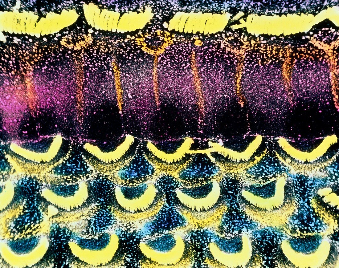
| Organ of Corti. Coloured scanning electron micrograph of hair cells (yellow) in the organ of Corti in the inner ear. They are divided into outer hair cells (v-shaped rows) and inner hair cells (top). Hair cells are immersed in a fluid called endolymph and react to the pressure of sound waves with an undulatory motion. This motion stimulates a nerve ending in each group of hair cells which carries a signal to various parts of the brain via the cochlear nerve. The two types of hair cells are divided by the pillar cells (pink) seen here covered by microvilli (speckles). Magnification: x1,320 at 6x7cm size. x1570 at 4x5 inch size | |
| Licence : | Droits gérés |
| Crédit: | Science Photo Library / PHOTO INSOLITE REALITE & V. GREMET |
| Taille de l’image : | 4724 px × 3737 px |
| Model Release : | Non requis |
| Property Release : | Non requis |
| Restrictions : | - |
Prix pour cette image À partir de 45 €
Produit vendu
(Calendrier, Carte postale, Carte de vœux, Impression sur textile, Packaging etc)
À partir de 45 €
Usage commercial
(Affichage, Annonce presse, Annonce TV, Carte, Digital - hors rés. sociaux, Digital - rés. sociaux etc)
À partir de 45 €
Éditorial
(Digital, Journal, Livre, Livre pratique, Magazine, Télévision etc)
À partir de 60 €
Usage non-commercial
(Digital - hors rés. sociaux, Digital - rés. sociaux etc)
À partir de 120 €