False-colour SEM of ciliated hair cells in the ear
Numéro d’image : 11872122
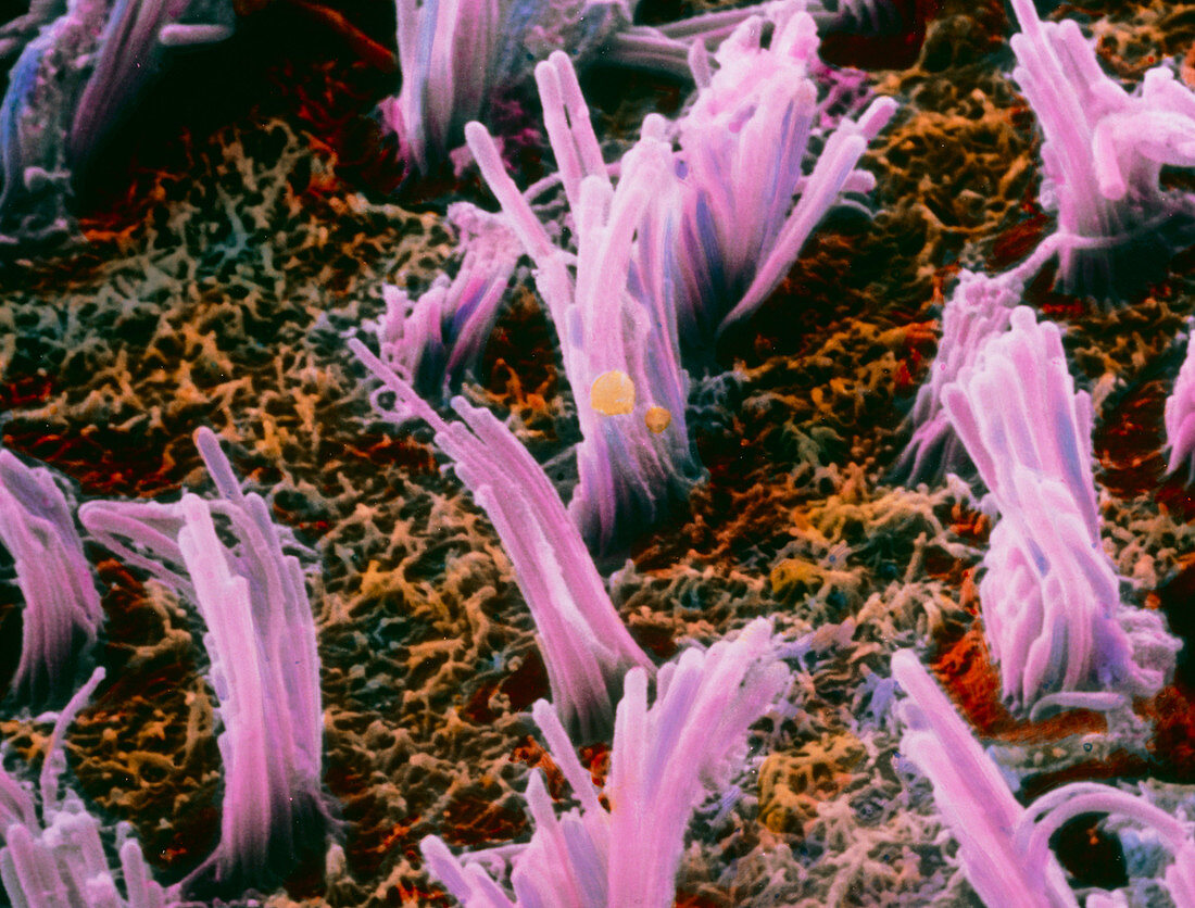
| Inner ear. False-colour scanning electron micrograph of bundles of ciliated hair cells (pink) situated in the macula utriculi within the human inner ear. They are responsible for maintaining equilibrium and the detection of directional and positional movement such as the tilting of the head. These hair cells are immersed in a fluid,called the endolymph,and are connected to the vestibular nerve. Currents in this fluid displace the position of the hair cells resulting in the production of pulses which are sent to the brain via the vestibular nerve. Magnification: x3550 at 6x7cm size. x5580 at 4x5ins | |
| Licence : | Droits gérés |
| Crédit: | Science Photo Library / UNIVERSITY LA SAPIENZA, ROME / DEPT. OF ANATOMY / PROF. P. MOTTA |
| Taille de l’image : | 3588 px × 2728 px |
| Model Release : | Non requis |
| Property Release : | Non requis |
| Restrictions : | - |
Prix pour cette image À partir de 45 €
Produit vendu
(Calendrier, Carte postale, Carte de vœux, Impression sur textile, Packaging etc)
À partir de 45 €
Usage commercial
(Affichage, Annonce presse, Annonce TV, Carte, Digital - hors rés. sociaux, Digital - rés. sociaux etc)
À partir de 45 €
Éditorial
(Digital, Journal, Livre, Livre pratique, Magazine, Télévision etc)
À partir de 60 €
Usage non-commercial
(Digital - hors rés. sociaux, Digital - rés. sociaux etc)
À partir de 120 €