F/col SEM of hair cells on organ of Corti
Numéro d’image : 11872119
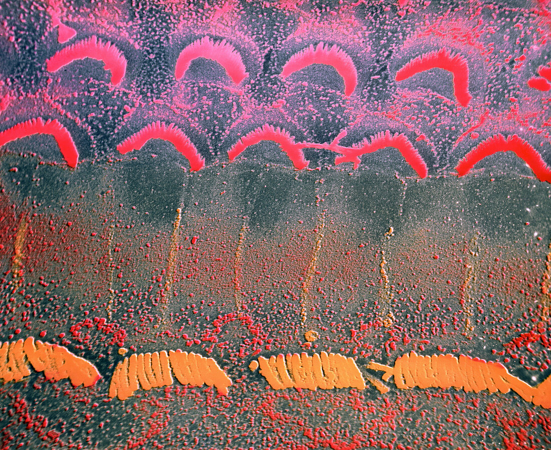
| False-colour scanning electron micrograph (SEM) of the surface of the organ of Corti in the human inner ear,following the removal of the covering,tectorial membrane. At bottom is a single row of inner hair cells containing stereocilia (hair- like projections) that are linearly arranged. In contrast,the stereocilia associated with the upper layers of outer hair cells are arranged in an inverted V-shape. Each hair cell may support up to 100 individual stereocilia,which function in translating the movement caused by sound waves into electrical impulses that are transmitted to the brain via the cochlear nerve. Magnification: x730 at 35mm,x1460 at 6x7cm size | |
| Licence : | Droits gérés |
| Crédit: | Science Photo Library / PHOTO INSOLITE REALITE & V. GREMET |
| Taille de l’image : | 4224 px × 3448 px |
| Model Release : | Non requis |
| Property Release : | Non requis |
| Restrictions : | - |
Prix pour cette image À partir de 45 €
Produit vendu
(Calendrier, Carte postale, Carte de vœux, Impression sur textile, Packaging etc)
À partir de 45 €
Usage commercial
(Affichage, Annonce presse, Annonce TV, Carte, Digital - hors rés. sociaux, Digital - rés. sociaux etc)
À partir de 45 €
Éditorial
(Digital, Journal, Livre, Livre pratique, Magazine, Télévision etc)
À partir de 60 €
Usage non-commercial
(Digital - hors rés. sociaux, Digital - rés. sociaux etc)
À partir de 120 €