Cone cell contents
Numéro d’image : 11871954
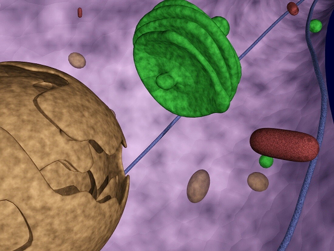
| Cone cell. Computer model of the contents of a cell body of a cone nerve cell from the human retina. The retina is the light-sensitive tissue that lines the inside of the eye. Each cone can detect either red,blue or green light,converting it into a nervous impulse that travels to the optic nerve and on to the brain. The cell nucleus (brown) is at bottom left. The Golgi body (green) completes the assembly of any proteins that will be secreted,stored or incorporated into the cell membrane. This image was created at the Laboratory of Neuro Imaging (LONI) at UCLA,USA | |
| Licence : | Droits gérés |
| Crédit: | Science Photo Library / Toga, Arthur / UCLA |
| Taille de l’image : | 3150 px × 2363 px |
| Model Release : | Non requis |
| Property Release : | Non requis |
| Restrictions : | - |
Prix pour cette image À partir de 45 €
Produit vendu
(Calendrier, Carte postale, Carte de vœux, Impression sur textile, Packaging etc)
À partir de 45 €
Usage commercial
(Affichage, Annonce presse, Annonce TV, Carte, Digital - hors rés. sociaux, Digital - rés. sociaux etc)
À partir de 45 €
Éditorial
(Digital, Journal, Livre, Livre pratique, Magazine, Télévision etc)
À partir de 60 €
Usage non-commercial
(Digital - hors rés. sociaux, Digital - rés. sociaux etc)
À partir de 120 €
Mots clés
- Amérique,
- anatomie,
- apparatus,
- appareil,
- cellule de cône,
- corps cellulaire,
- corps de Golgi,
- corps humain,
- Etats-Unis,
- Golgi,
- L.A.,
- LA,
- Loni,
- Los Angeles,
- modèle d'ordinateur,
- N/A,
- nerfs,
- neurone,
- noyau,
- nucleus,
- oeil,
- retina,
- rétine,
- sens,
- sens visuel,
- système nerveux,
- type d'ordinateur,
- UCLA,
- université de Californie,
- US,
- USA,
- vision,
- vue