Light micrograph of a section through the eye wall
Numéro d’image : 11871927
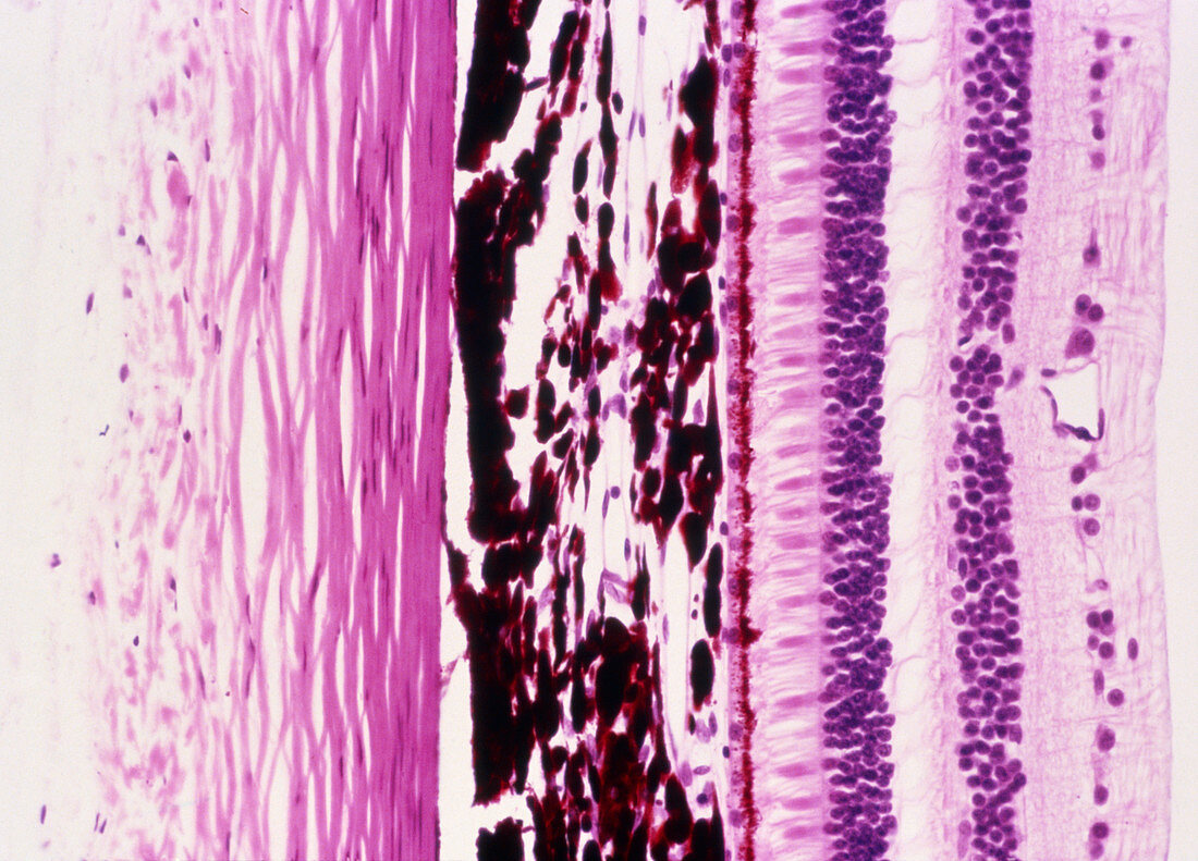
| Eye wall. Light micrograph of a cross section through the eye wall. The outer layer (left,pink) is the sclera,connective tissue arranged in bundles. The next layer (centre,dark red) is the choroid. This layer contains blood vessels and a pigment to absorb excess light to prevent blurred vision. Inside of the choroid is the multi-layered retina. Its first layer is a line of pigment cells (red),immediately followed by the light sensitive rod and cone cells (pink). For light to reach these cells it must pass through layers of nerve cells. The nerve cell nuclei are visible as layers of purple dots. Haematoxylin and eosin stained. Magnification x100 at 35mm size | |
| Licence : | Droits gérés |
| Crédit: | Science Photo Library / Biophoto Associates |
| Taille de l’image : | 4924 px × 3543 px |
| Model Release : | Non requis |
| Property Release : | Non requis |
| Restrictions : | - |
Prix pour cette image À partir de 45 €
Produit vendu
(Calendrier, Carte postale, Carte de vœux, Impression sur textile, Packaging etc)
À partir de 45 €
Usage commercial
(Affichage, Annonce presse, Annonce TV, Carte, Digital - hors rés. sociaux, Digital - rés. sociaux etc)
À partir de 45 €
Éditorial
(Digital, Journal, Livre, Livre pratique, Magazine, Télévision etc)
À partir de 60 €
Usage non-commercial
(Digital - hors rés. sociaux, Digital - rés. sociaux etc)
À partir de 120 €
Mots clés
- anatomie,
- choroïde,
- corps humain,
- mur d'œil,
- oeil,
- retina,
- rétine,
- sclera,
- sclère,
- sens visuel,
- slérotique,
- structure,
- tache,
- vision,
- vue