Coloured SEM of iris cells of the eye,& monocyte
Numéro d’image : 11871921
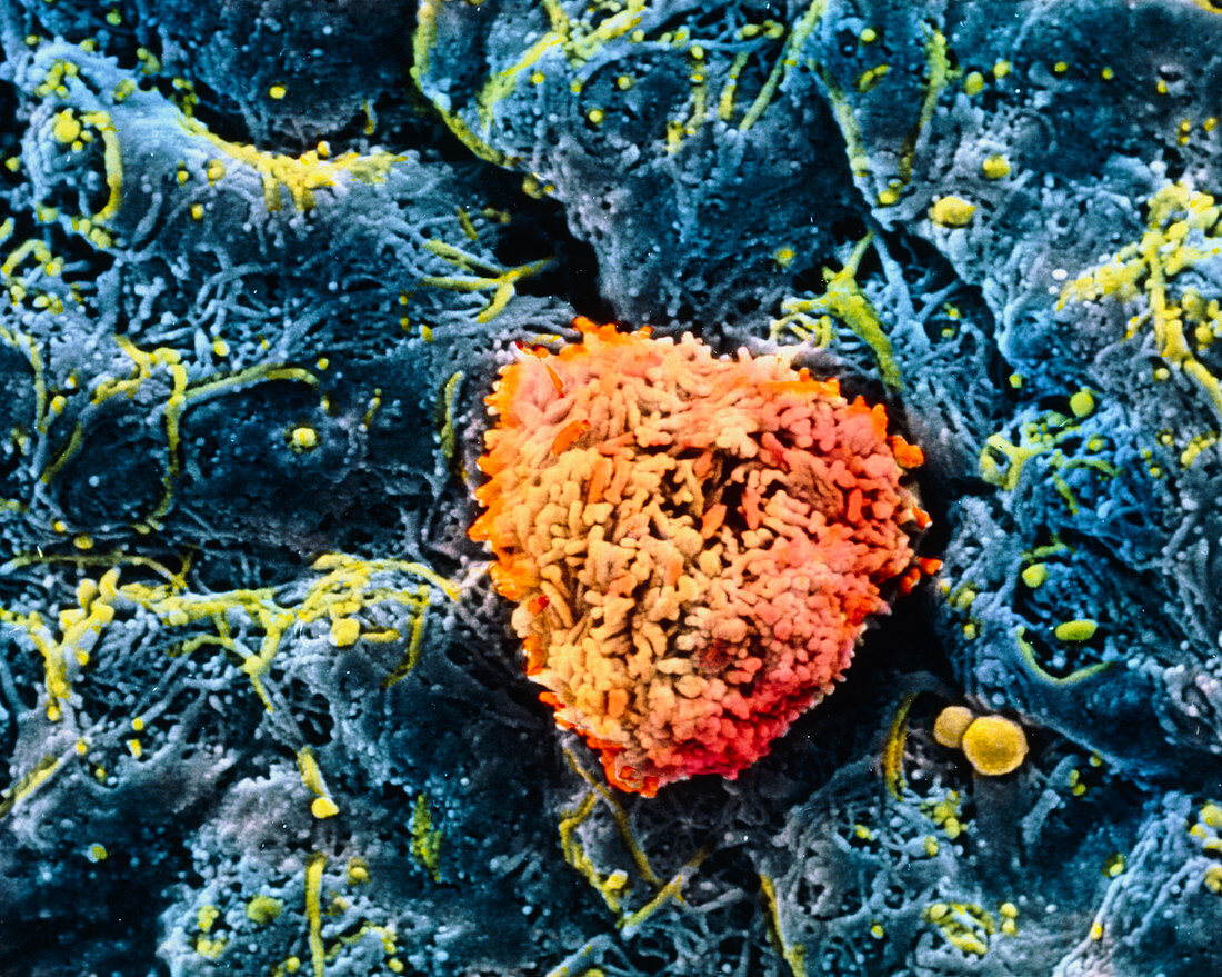
| Iris cells of the eye. Coloured Scanning Electron Micrograph (SEM) of cells of the anterior surface of the iris. The iris forms the delicate and adjustable diaphragm which surrounds the pupil. It is irregular and rough,its surface composed of fibroblast cells (blue). These cells are joined loosely by connective tissue fibres (green). Granules (green) may be pigments escaped from pigment cells (melanocytes) beneath this layer. A monocyte (orange) is the largest type of white blood cell and serves an immune function. The iris is movable,allowing light to enter the pupil. Magnification: x2,200 at 6x7cm size. x2,860 at 4x5ins | |
| Licence : | Droits gérés |
| Crédit: | Science Photo Library / UNIVERSITY LA SAPIENZA, ROME / DEPT. OF ANATOMY / PROF. P. MOTTA |
| Taille de l’image : | 5005 px × 3999 px |
| Model Release : | Non requis |
| Property Release : | Non requis |
| Restrictions : | - |
Prix pour cette image À partir de 45 €
Produit vendu
(Calendrier, Carte postale, Carte de vœux, Impression sur textile, Packaging etc)
À partir de 45 €
Usage commercial
(Affichage, Annonce presse, Annonce TV, Carte, Digital - hors rés. sociaux, Digital - rés. sociaux etc)
À partir de 45 €
Éditorial
(Digital, Journal, Livre, Livre pratique, Magazine, Télévision etc)
À partir de 60 €
Usage non-commercial
(Digital - hors rés. sociaux, Digital - rés. sociaux etc)
À partir de 120 €