False-colour SEM of the cell surface of the cornea
Numéro d’image : 11871913
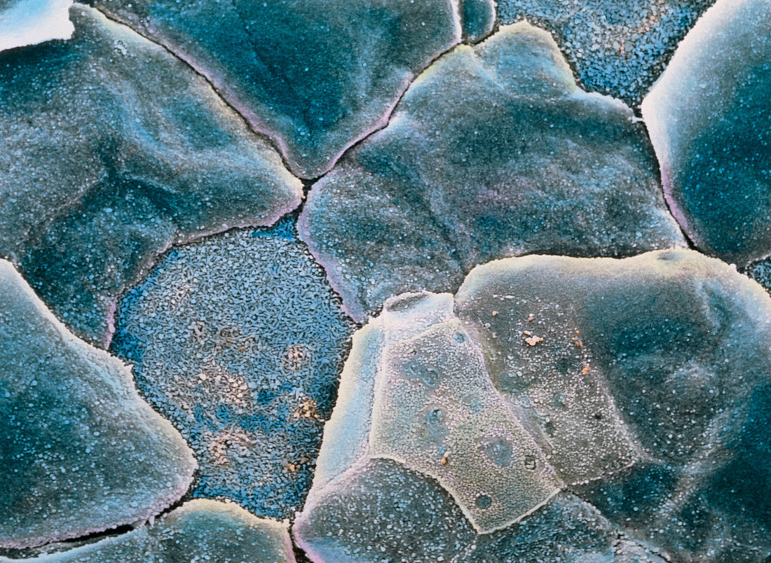
| False-colour scanning electron micrograph (SEM) of the cell surface of the cornea of the eye. These cells are flattened,closely packed,and trans- parent. This is a stratified squamous epithelium 5 cell layers deep. Cells at centre left and upper right have peeled away,to reveal tiny ridges (microplicae) on the inner surface which bind each cell together. Although rich in nerves,there are no blood vessels in the cornea: this would affect its transparency. This external surface over the anterior of the eye has a convex curve to provide the main mechanism for focusing light onto the retina. Magnification: x1170 at 6x7cm size. Magnification: x1800 at 4x5 inch size | |
| Licence : | Droits gérés |
| Crédit: | Science Photo Library / UNIVERSITY LA SAPIENZA, ROME / DEPT. OF ANATOMY / PROF. P. MOTTA |
| Taille de l’image : | 3726 px × 2723 px |
| Model Release : | Non requis |
| Property Release : | Non requis |
| Restrictions : | - |
Prix pour cette image À partir de 45 €
Produit vendu
(Calendrier, Carte postale, Carte de vœux, Impression sur textile, Packaging etc)
À partir de 45 €
Usage commercial
(Affichage, Annonce presse, Annonce TV, Carte, Digital - hors rés. sociaux, Digital - rés. sociaux etc)
À partir de 45 €
Éditorial
(Digital, Journal, Livre, Livre pratique, Magazine, Télévision etc)
À partir de 60 €
Usage non-commercial
(Digital - hors rés. sociaux, Digital - rés. sociaux etc)
À partir de 120 €