False-colour SEM of the cell surface of the cornea
Numéro d’image : 11871912
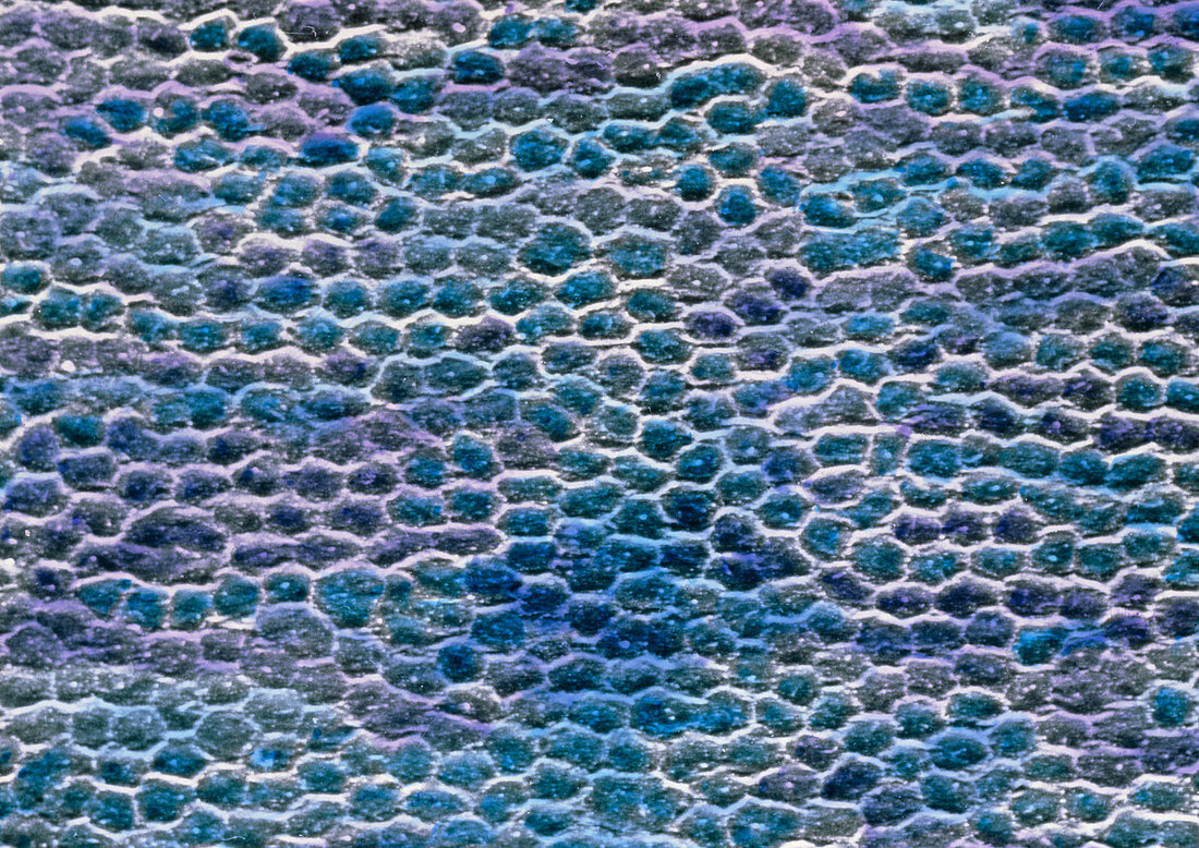
| False-colour scanning electron micrograph (SEM) of the surface of the cornea of the eye. The matrix- like pattern (seen here) consists of individual flattened transparent cells. This is a stratified squamous epithelium which is 5 cell layers deep. Tiny ridges (unseen) on each cell surface helps bind these cells together. Although richly supplied with nerves,there are no blood vessels in the cornea: this would affect its transparency. This external surface which encloses the anterior of the eye has a fixed convex curve; it provides the main mechanism for focusing light images onto the retina. Magnification: x90 at 6x7cm size. Magnification: x140 at 4x5 inch size | |
| Licence : | Droits gérés |
| Crédit: | Science Photo Library / UNIVERSITY LA SAPIENZA, ROME / DEPT. OF ANATOMY / PROF. P. MOTTA |
| Taille de l’image : | 4841 px × 3425 px |
| Model Release : | Non requis |
| Property Release : | Non requis |
| Restrictions : | - |
Prix pour cette image À partir de 45 €
Produit vendu
(Calendrier, Carte postale, Carte de vœux, Impression sur textile, Packaging etc)
À partir de 45 €
Usage commercial
(Affichage, Annonce presse, Annonce TV, Carte, Digital - hors rés. sociaux, Digital - rés. sociaux etc)
À partir de 45 €
Éditorial
(Digital, Journal, Livre, Livre pratique, Magazine, Télévision etc)
À partir de 60 €
Usage non-commercial
(Digital - hors rés. sociaux, Digital - rés. sociaux etc)
À partir de 120 €