Fundus camera image of a normal retina,Caucasian
Numéro d’image : 11871893
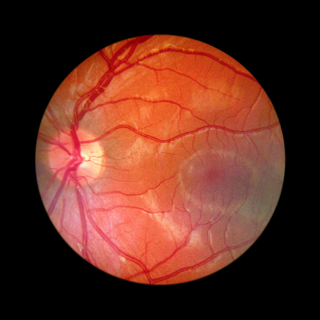
| Fundus camera image of the retina of a normal eye,showing the distribution of the retinal veins & arteries: the central retinal artery (a branch of the opthalmic artery) enters the optic nerve 2 cm before it reaches the eyeball,emerging through the retina in the centre of the optic disc (the blind spot of the eye,the pale area on the left). The dark oval region free of large blood vessels (right) is the macula,in the centre of which the fovea is found. The fovea is the region with highest visual acuity. An extension of the optic nerve,the retina consists of photosensitive cells (rods & cones) which translate light energy into nervous impulses | |
| Licence : | Droits gérés |
| Crédit: | Science Photo Library / McClenaghan, Rory |
| Taille de l’image : | 4724 px × 4724 px |
| Model Release : | Non requis |
| Property Release : | Non requis |
| Restrictions : | - |
Prix pour cette image À partir de 45 €
Produit vendu
(Calendrier, Carte postale, Carte de vœux, Impression sur textile, Packaging etc)
À partir de 45 €
Usage commercial
(Affichage, Annonce presse, Annonce TV, Carte, Digital - hors rés. sociaux, Digital - rés. sociaux etc)
À partir de 45 €
Éditorial
(Digital, Journal, Livre, Livre pratique, Magazine, Télévision etc)
À partir de 60 €
Usage non-commercial
(Digital - hors rés. sociaux, Digital - rés. sociaux etc)
À partir de 120 €