Fundus camera image of a normal retina,Asian
Numéro d’image : 11871887
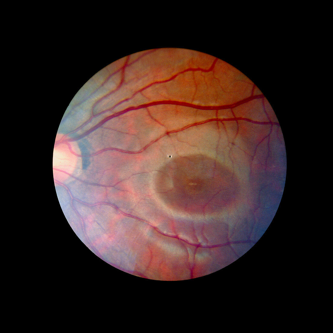
| Fundus camera image of the retina of a normal eye (Asian),showing the retinal veins & arteries emerging from the optic disc (the blind spot of the eye,pale area on left). The dark oval free of large blood vessels (right) is the macula,in the centre of which is the fovea. The fovea has the highest density of cones (photosensitive cells which function best in bright light),& therefore greatest acuity of vision. The black dot in the centre of the image may be the remnant of the hyaloid artery which supplied blood to the lens during embryonic development. An extension of the optic nerve,the retina consists of cells which translate light energy into nervous impulses | |
| Licence : | Droits gérés |
| Crédit: | Science Photo Library / McClenaghan, Rory |
| Taille de l’image : | 4724 px × 4724 px |
| Model Release : | Non requis |
| Property Release : | Non requis |
| Restrictions : | - |
Prix pour cette image À partir de 45 €
Produit vendu
(Calendrier, Carte postale, Carte de vœux, Impression sur textile, Packaging etc)
À partir de 45 €
Usage commercial
(Affichage, Annonce presse, Annonce TV, Carte, Digital - hors rés. sociaux, Digital - rés. sociaux etc)
À partir de 45 €
Éditorial
(Digital, Journal, Livre, Livre pratique, Magazine, Télévision etc)
À partir de 60 €
Usage non-commercial
(Digital - hors rés. sociaux, Digital - rés. sociaux etc)
À partir de 120 €