Ciliary body and iris of the eye
Numéro d’image : 11871880
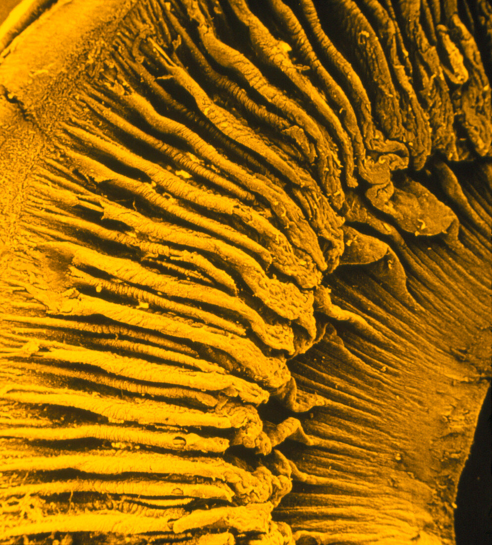
| False-colour,low-power scanning electron micrograph (SEM) of the human eye,showing the ciliary body & iris. The deeply folded tissue at left & top is the ciliary body,which normally encircles & controls the lens of the eye,flattening it to focus distant objects onto the retina & making it more spherical to focus closer ones. In this view,seen from the back of the eye,the lens has been removed. As a result,one can see through to the less folded muscles of the iris,which control the aperture of the pupil (the black area at bottom right) & so the amount of light that reaches the retina. Magnification: x2.5 at 35mm size. Yellow tint. Reference: MICROCOSMOS,figure 2.9,page 19 | |
| Licence : | Droits gérés |
| Crédit: | Science Photo Library / CNRI |
| Taille de l’image : | 2598 px × 2877 px |
| Model Release : | Non requis |
| Property Release : | Non requis |
| Restrictions : | - |
Prix pour cette image À partir de 45 €
Produit vendu
(Calendrier, Carte postale, Carte de vœux, Impression sur textile, Packaging etc)
À partir de 45 €
Usage commercial
(Affichage, Annonce presse, Annonce TV, Carte, Digital - hors rés. sociaux, Digital - rés. sociaux etc)
À partir de 45 €
Éditorial
(Digital, Journal, Livre, Livre pratique, Magazine, Télévision etc)
À partir de 60 €
Usage non-commercial
(Digital - hors rés. sociaux, Digital - rés. sociaux etc)
À partir de 120 €