LM through section of human eye
Numéro d’image : 11871872
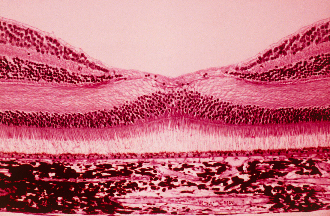
| Light micrograph through a section of the human eye in the region of the fovea,a depression in the photosensitive retina,the inner lining of the eye. The foveal retina is the area of greatest visual acuity and contains only cone receptor cells (the hump-backed layers). N.B. The retina is composed of 2 types of photosensitive cells,rods & cones. The rods differentiate light of differing intensity (analagous to a black & white image); cone cells are of 3 types receptive to blue,green & red light & form a system for colour perception. The broad bottom layer in image is the choroid layer,supporting & separated from the retina by a layer of pigmented epithelial cells. Magnification: x300 when printed at 10 centimetres wide | |
| Licence : | Droits gérés |
| Crédit: | Science Photo Library / Cox, Gene |
| Taille de l’image : | 4981 px × 3267 px |
| Model Release : | Non requis |
| Property Release : | Non requis |
| Restrictions : | - |
Prix pour cette image À partir de 45 €
Produit vendu
(Calendrier, Carte postale, Carte de vœux, Impression sur textile, Packaging etc)
À partir de 45 €
Usage commercial
(Affichage, Annonce presse, Annonce TV, Carte, Digital - hors rés. sociaux, Digital - rés. sociaux etc)
À partir de 45 €
Éditorial
(Digital, Journal, Livre, Livre pratique, Magazine, Télévision etc)
À partir de 60 €
Usage non-commercial
(Digital - hors rés. sociaux, Digital - rés. sociaux etc)
À partir de 120 €