Fundus camera image of a normal retina,Asian
Numéro d’image : 11871594
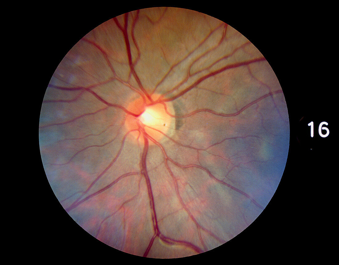
| Fundus camera image of the retina of a normal eye,showing the distribution of the retinal veins & arteries. The central retinal artery enters the optic nerve before it reaches the eyeball,and emerges from the centre of the optic disc (the blind spot of the eye,the pale central area). On the right can be seen the dark edge of the macula,a region free of large blood vessels where the fovea (the area of highest visual acuity) is found. An extension of the optic nerve,the retina consists of photosensitive cells (rods & cones) which translate light energy into nervous impulses. This retina has a greenish hue because the subject was Asian. In Caucasians it is reddish | |
| Licence : | Droits gérés |
| Crédit: | Science Photo Library / McClenaghan, Rory |
| Taille de l’image : | 3543 px × 2766 px |
| Model Release : | Non requis |
| Property Release : | Non requis |
| Restrictions : | - |
Prix pour cette image À partir de 45 €
Produit vendu
(Calendrier, Carte postale, Carte de vœux, Impression sur textile, Packaging etc)
À partir de 45 €
Usage commercial
(Affichage, Annonce presse, Annonce TV, Carte, Digital - hors rés. sociaux, Digital - rés. sociaux etc)
À partir de 45 €
Éditorial
(Digital, Journal, Livre, Livre pratique, Magazine, Télévision etc)
À partir de 60 €
Usage non-commercial
(Digital - hors rés. sociaux, Digital - rés. sociaux etc)
À partir de 120 €