Nerve cell growth
Numéro d’image : 11871291
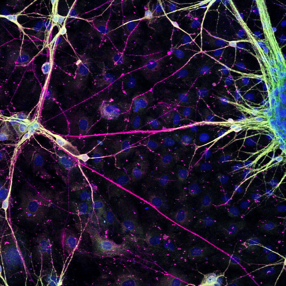
| Nerve cell growth. Light micrograph of nerve cells (neurons) with immunofluorescent staining. These cells have been grown in culture. The stains show neurites (thin strands,either axons or dendrites) connecting nerve cell bodies. Short neurites are green,long ones are pink. Nerve cell bodies are blue,but non-neuronal cell nuclei (also blue) are seen as well. Neurons transmit electrical signals around the body,especially the brain and the spinal cord. These neurons have been grown from NT2 cells,a human teratocarcinoma cell line capable of differentiation into nerve cells. Such research may make neural regeneration possible for treating spinal paralysis and nerve disorders | |
| Licence : | Droits gérés |
| Crédit: | Science Photo Library / Paquet-Durand, Francois |
| Taille de l’image : | 2511 px × 2511 px |
| Model Release : | Non requis |
| Property Release : | Non requis |
| Restrictions : | - |
Prix pour cette image À partir de 45 €
Produit vendu
(Calendrier, Carte postale, Carte de vœux, Impression sur textile, Packaging etc)
À partir de 45 €
Usage commercial
(Affichage, Annonce presse, Annonce TV, Carte, Digital - hors rés. sociaux, Digital - rés. sociaux etc)
À partir de 45 €
Éditorial
(Digital, Journal, Livre, Livre pratique, Magazine, Télévision etc)
À partir de 60 €
Usage non-commercial
(Digital - hors rés. sociaux, Digital - rés. sociaux etc)
À partir de 120 €
Mots clés
- axone,
- axones,
- biologie,
- biologique,
- cellule,
- cellules nerveuses,
- colorer,
- corps,
- cultivé,
- culture,
- dendrite,
- dendrites,
- immunofluorescence,
- microscope optique,
- microscopie optique,
- nerf,
- neurité,
- neurites,
- neurologie,
- neurone,
- neurones,
- neuroscience,
- noyau,
- nucleus,
- sali,
- souillé,
- système nerveux,
- taché