LM of cerebellar tissue with Purkinje cel
Numéro d’image : 11871115
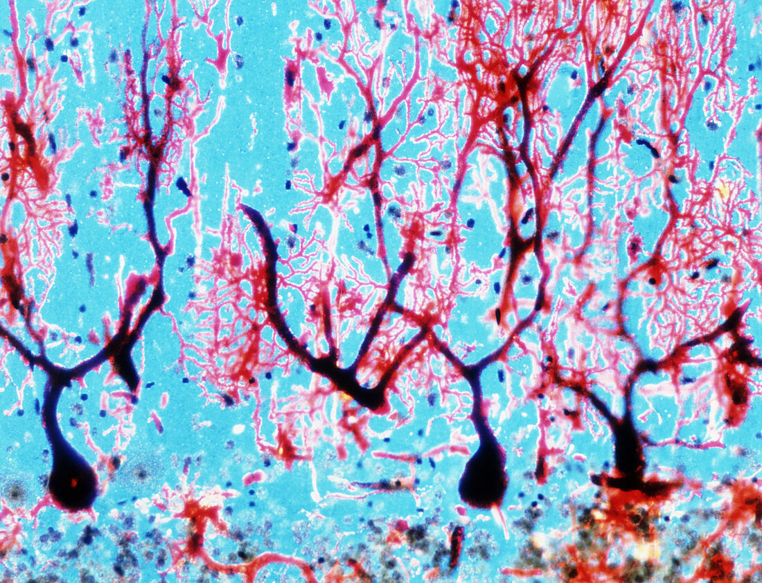
| Purkinje nerve cells. Light micrograph of a cross section of cerebellar tissue showing the highly differentiated brain cells known as Purkinje cells. They are formed by a flask-shaped body (dark at bottom) from which dense (dark red) dendritic ramifications depart. The Purkinje cells are found in the cerebellar cortex at the junction between the molecular layer,which occupies most of the frame,and the granular layer just visible at bottom. This contains a huge number of microneurones (3-7 millions per cubic millimetre),known as granule cells (round blue-grey). Magnification: x60 at 6x4.5cm size | |
| Licence : | Droits gérés |
| Crédit: | Science Photo Library / Pasieka, Alfred |
| Taille de l’image : | 4153 px × 3176 px |
| Model Release : | Non requis |
| Property Release : | Non requis |
| Restrictions : | - |
Prix pour cette image À partir de 45 €
Produit vendu
(Calendrier, Carte postale, Carte de vœux, Impression sur textile, Packaging etc)
À partir de 45 €
Usage commercial
(Affichage, Annonce presse, Annonce TV, Carte, Digital - hors rés. sociaux, Digital - rés. sociaux etc)
À partir de 45 €
Éditorial
(Digital, Journal, Livre, Livre pratique, Magazine, Télévision etc)
À partir de 60 €
Usage non-commercial
(Digital - hors rés. sociaux, Digital - rés. sociaux etc)
À partir de 120 €