Nerve cells
Numéro d’image : 11871108
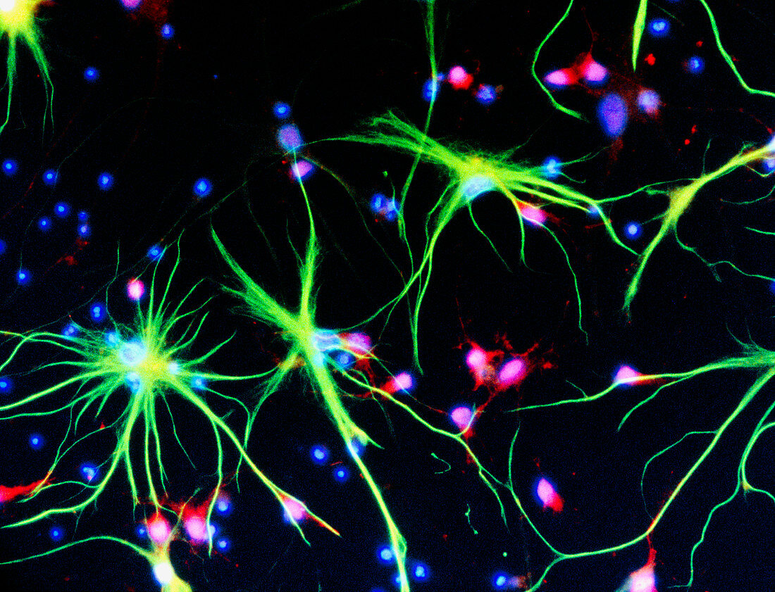
| Immunofluorescent Light Micrograph of neuron cells and astrocytes in mammalian spinal cord. Here,neuron cells stain red: the cell body appears pink,with nerve fibres faintly seen which are the route of transmission of nerve impulses. In the foreground are astrocyte cells (green): star- shaped connective tissue cells which provide support and nutrients for the neurons. It is thought that astrocytes may take part in infor- mation storage. Blue dots are the nuclei of other support cells. Immunofluorescence is a staining technique which uses antibodies to attach fluores- cent dyes to specific tissues and molecules in the cell. Magnification x400 at 5x7cm,x375 at 6x4.5cm x200 at 35mm | |
| Licence : | Droits gérés |
| Crédit: | Science Photo Library / Kedersha, Nancy / UCLA |
| Taille de l’image : | 5197 px × 3974 px |
| Model Release : | Non requis |
| Property Release : | Non requis |
| Restrictions : | - |
Prix pour cette image À partir de 45 €
Produit vendu
(Calendrier, Carte postale, Carte de vœux, Impression sur textile, Packaging etc)
À partir de 45 €
Usage commercial
(Affichage, Annonce presse, Annonce TV, Carte, Digital - hors rés. sociaux, Digital - rés. sociaux etc)
À partir de 45 €
Éditorial
(Digital, Journal, Livre, Livre pratique, Magazine, Télévision etc)
À partir de 60 €
Usage non-commercial
(Digital - hors rés. sociaux, Digital - rés. sociaux etc)
À partir de 120 €