False-colour SEM of a bundle of motor nerve fibres
Numéro d’image : 11870988
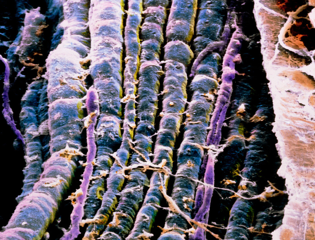
| Nerve fibres. False-colour scanning electron micrograph of a bundle of motor nerve fibres (blue). The bundle as a whole is surrounded by a cylindrical sheath of connective tissue,also known as the epineurium,which is partly visible at extreme right (soft brown). The epineurium has been opened to show the nerve fibres and a few capillaries (violet). Each fibre is surrounded by a thick myelin layer,mostly formed by lipid material,which derives from the Schwann cells. Through these fibres messages are sent from the brain to the muscles with a maximum speed of 100m per second. Magnification: x 500 at 6x7cm size. x780 at 4x5ins | |
| Licence : | Droits gérés |
| Crédit: | Science Photo Library / UNIVERSITY LA SAPIENZA, ROME / DEPT. OF ANATOMY / PROF. P. MOTTA |
| Taille de l’image : | 4800 px × 3661 px |
| Model Release : | Non requis |
| Property Release : | Non requis |
| Restrictions : | - |
Prix pour cette image À partir de 45 €
Produit vendu
(Calendrier, Carte postale, Carte de vœux, Impression sur textile, Packaging etc)
À partir de 45 €
Usage commercial
(Affichage, Annonce presse, Annonce TV, Carte, Digital - hors rés. sociaux, Digital - rés. sociaux etc)
À partir de 45 €
Éditorial
(Digital, Journal, Livre, Livre pratique, Magazine, Télévision etc)
À partir de 60 €
Usage non-commercial
(Digital - hors rés. sociaux, Digital - rés. sociaux etc)
À partir de 120 €