TEM of the myelin sheath around the auditory nerve
Numéro d’image : 11870985
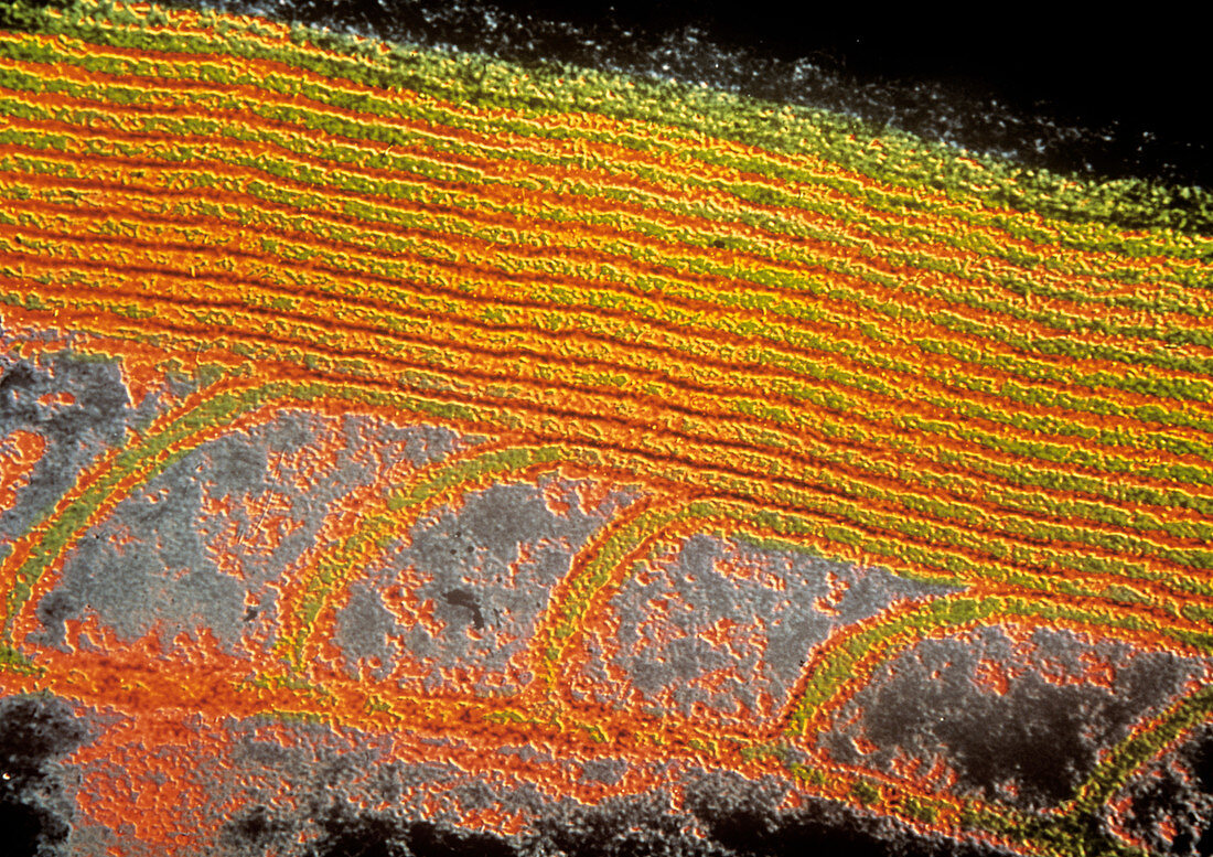
| False-colour transmission electron micrograph (TEM) showing the myelin sheath surrounding the human auditory nerve. In longitudinal section,concentric layers of myelin sheath appear as orange bands at top of image. The layers are formed by envelopes of the cytoplasm of schwann cells (specialised cells which envelope all axons support). Myelination begins during foetal development with the introduction of a mesaxon (invagination of single nerve fibre into schwann cell) which wraps around the axon (evident towards bottom of image - blue area is axon). Magnification: X 88,000 at 35mm size. N.B. In all nerve fibres,the rate of conduction | |
| Licence : | Droits gérés |
| Crédit: | Science Photo Library / CNRI |
| Taille de l’image : | 4989 px × 3525 px |
| Model Release : | Non requis |
| Property Release : | Non requis |
| Restrictions : | - |
Prix pour cette image À partir de 45 €
Produit vendu
(Calendrier, Carte postale, Carte de vœux, Impression sur textile, Packaging etc)
À partir de 45 €
Usage commercial
(Affichage, Annonce presse, Annonce TV, Carte, Digital - hors rés. sociaux, Digital - rés. sociaux etc)
À partir de 45 €
Éditorial
(Digital, Journal, Livre, Livre pratique, Magazine, Télévision etc)
À partir de 60 €
Usage non-commercial
(Digital - hors rés. sociaux, Digital - rés. sociaux etc)
À partir de 120 €