Spinal nerve tissue
Numéro d’image : 11870954
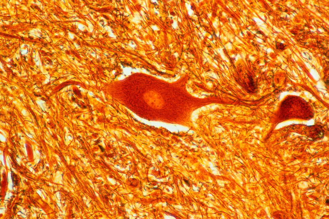
| Light micrograph of a motorneuron cell from the ventral horn of a human spinal cord,shown with Bodian staining. The pale central area is the nucleus,surrounded by the brown cell soma and extended dendrites. The soma and dendrites are the main recipient surfaces of the cell. The ventral horn of the spinal column is composed mainly of grey matter,in which motoneurons are found among large areas of connective tissue. Magnification x250 at 5x7cm,x125 at 35mm size | |
| Licence : | Droits gérés |
| Crédit: | Science Photo Library / Michler, Astrid & Hans-Frieder |
| Taille de l’image : | 4511 px × 3012 px |
| Model Release : | Non requis |
| Property Release : | Non requis |
| Restrictions : | - |
Prix pour cette image À partir de 45 €
Produit vendu
(Calendrier, Carte postale, Carte de vœux, Impression sur textile, Packaging etc)
À partir de 45 €
Usage commercial
(Affichage, Annonce presse, Annonce TV, Carte, Digital - hors rés. sociaux, Digital - rés. sociaux etc)
À partir de 45 €
Éditorial
(Digital, Journal, Livre, Livre pratique, Magazine, Télévision etc)
À partir de 60 €
Usage non-commercial
(Digital - hors rés. sociaux, Digital - rés. sociaux etc)
À partir de 120 €
Mots clés
- agrandissement,
- anatomie,
- anatomie humaine,
- central,
- corps,
- corps cellulaire,
- corps humain,
- dendrite,
- histologie,
- images,
- microscope optique,
- microscopie optique,
- moelle épinière,
- nerf,
- neurone,
- neurone moteur,
- péricaryon,
- photos au microscope,
- S.N.C.,
- SNC,
- soma,
- sujets,
- système nerveux,
- système nerveux central,
- tissus,
- vertébral