Coloured 3-D CT scan of a head with healt
Numéro d’image : 11870763
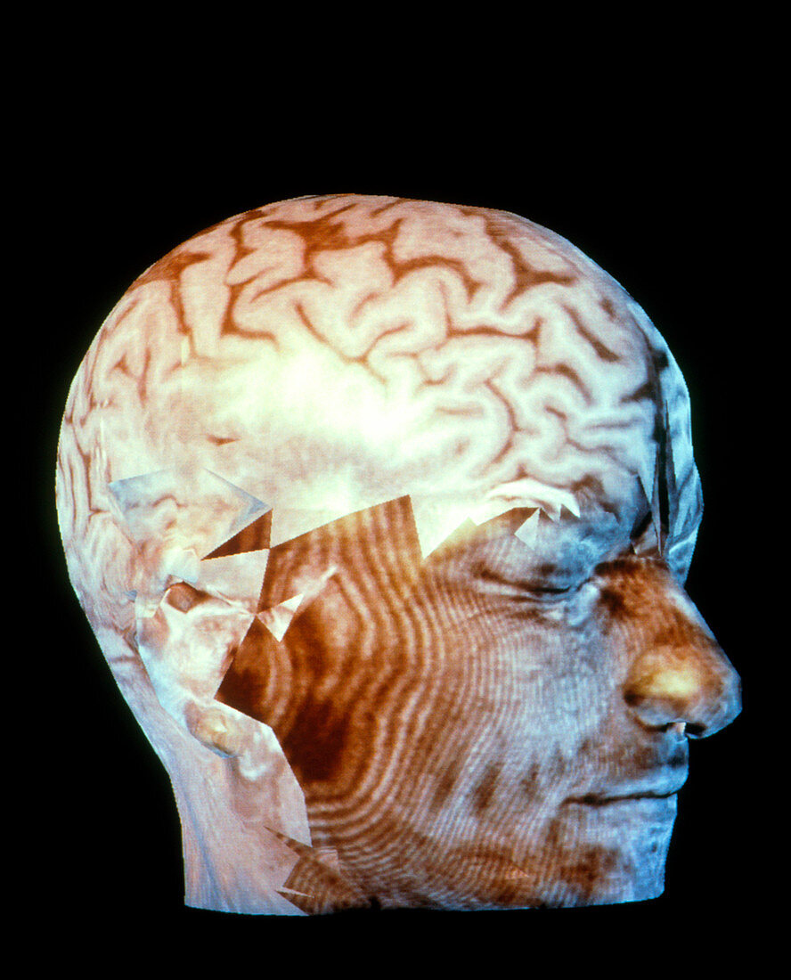
| Healthy brain. Coloured three-dimensional computed tomography (CT) scan of the side of a human head with a healthy brain projected onto it. The largest part of the brain,the folded cerebrum (at upper centre),is seen. This carries out conscious brain activity and is shown as a three-dimensional (3-D) magnetic resonance imaging (MRI) scan. CT scanning uses X-rays to produce 'slice' images of the body. Here,data from many 'slices' have been combined to create this 3-D view. The image was taken at Siemens Corporate Research Center in Princeton,USA. It was produced with new imaging software that enables 3-D scans to be freely rotated,aiding in medical diagnosis and surgery | |
| Licence : | Droits gérés |
| Crédit: | Science Photo Library / Steger, Volker |
| Taille de l’image : | 2860 px × 3543 px |
| Model Release : | Non requis |
| Property Release : | Non requis |
| Restrictions : | - |
Prix pour cette image À partir de 45 €
Produit vendu
(Calendrier, Carte postale, Carte de vœux, Impression sur textile, Packaging etc)
À partir de 45 €
Usage commercial
(Affichage, Annonce presse, Annonce TV, Carte, Digital - hors rés. sociaux, Digital - rés. sociaux etc)
À partir de 45 €
Éditorial
(Digital, Journal, Livre, Livre pratique, Magazine, Télévision etc)
À partir de 60 €
Usage non-commercial
(Digital - hors rés. sociaux, Digital - rés. sociaux etc)
À partir de 120 €