PET scan of brain of normal subject
Numéro d’image : 11870700
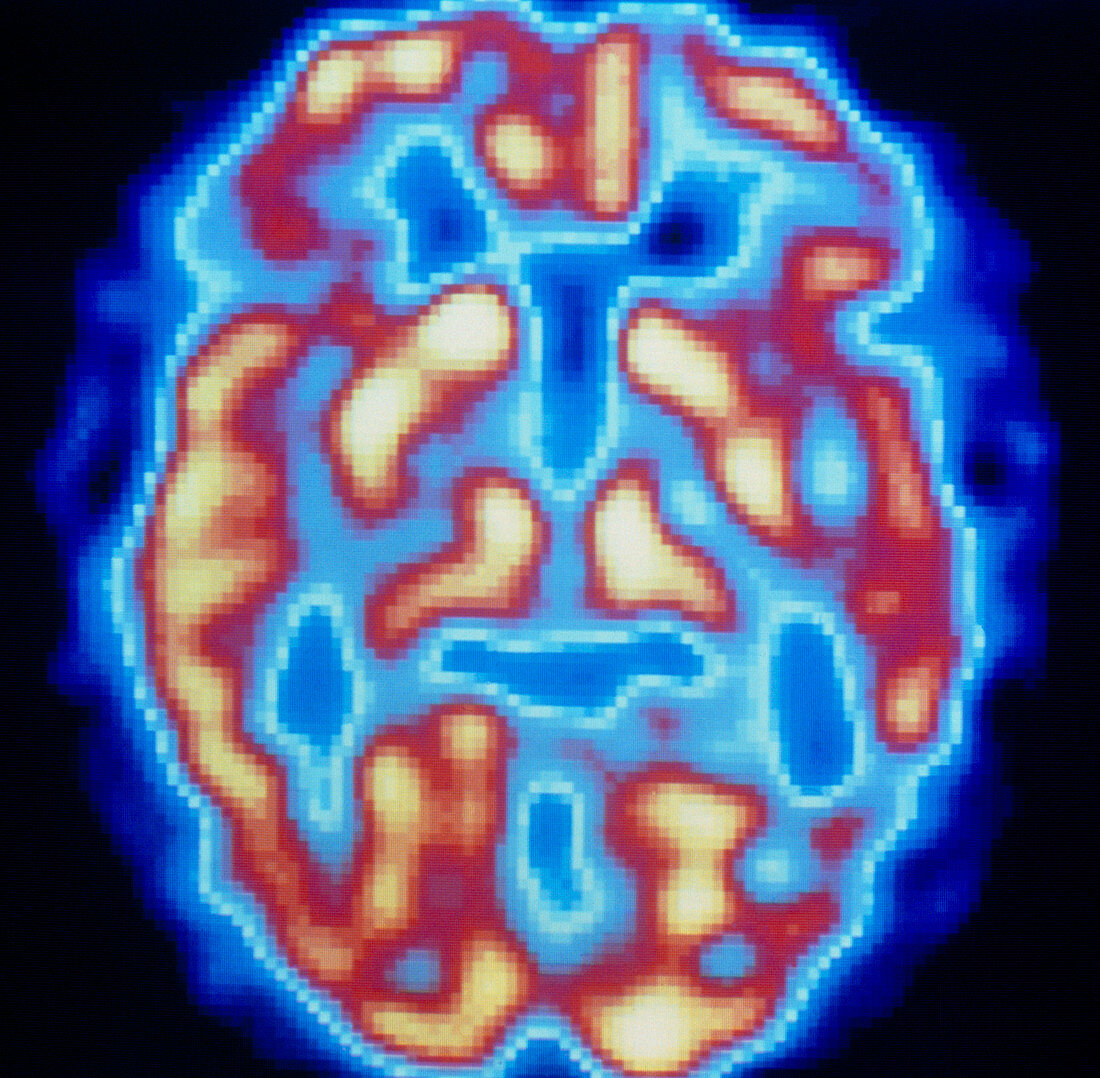
| Positron emission tomography (PET) scan of the brain (basal ganglia level) of a normal human subject. The colour-coded scan through this cerebral layer shows brain activity: from low (blue) to high (yellow). PET scanning relies on a radioactive tracer,injected into the bloodstream,to reveal metabolic activity in the brain. As seen here,normal brain metabolic activity produces a roughly symmetrical pattern in the yellow areas of left and right cerebral hemispheres. In many brain dysfunctions (such as Alzheimer's disease,epilepsy and schizophrenia),PET scans are useful in pinpointing specific brain areas which are metabolically affected by disease | |
| Licence : | Droits gérés |
| Crédit: | Science Photo Library / Beddow, Tim |
| Taille de l’image : | 4265 px × 4181 px |
| Model Release : | Non requis |
| Property Release : | Non requis |
| Restrictions : | - |
Prix pour cette image À partir de 45 €
Produit vendu
(Calendrier, Carte postale, Carte de vœux, Impression sur textile, Packaging etc)
À partir de 45 €
Usage commercial
(Affichage, Annonce presse, Annonce TV, Carte, Digital - hors rés. sociaux, Digital - rés. sociaux etc)
À partir de 45 €
Éditorial
(Digital, Journal, Livre, Livre pratique, Magazine, Télévision etc)
À partir de 60 €
Usage non-commercial
(Digital - hors rés. sociaux, Digital - rés. sociaux etc)
À partir de 120 €