NMR image showing structures of the brain
Numéro d’image : 11870690
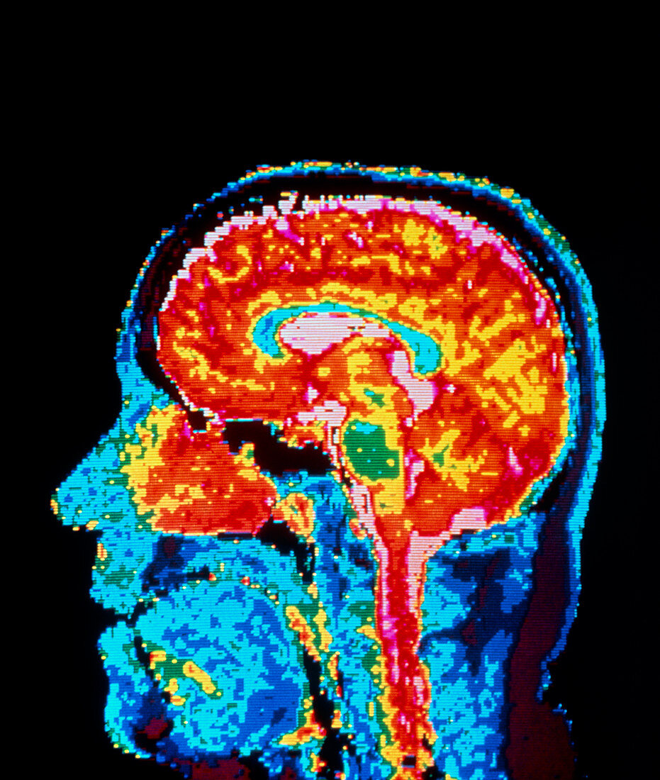
| False-colour nuclear magnetic resonance (NMR) image of a sagittal section through the human head,showing structures of a normal brain. The cerebrum (the 2 cerebral hemispheres) which forms the bulk of brain tissue appears orange-red. The pink areas at the centre,top,and surrounding the spinal cord at bottom represent cerebro-spinal fluid (CSF); the central pink area shows CSF in the ventricles (chambers) at the centre of the brain. The curved blue area surrounding the ventricles is the corpus callosum,a structure connecting the 2 cerebral hemispheres. At bottom right (top right of spinal cord) is the cerebell- um,the centre of balance & muscular coordination | |
| Licence : | Droits gérés |
| Crédit: | Science Photo Library |
| Taille de l’image : | 4200 px × 4961 px |
| Model Release : | Non requis |
| Property Release : | Non requis |
| Restrictions : | - |
Prix pour cette image À partir de 45 €
Produit vendu
(Calendrier, Carte postale, Carte de vœux, Impression sur textile, Packaging etc)
À partir de 45 €
Usage commercial
(Affichage, Annonce presse, Annonce TV, Carte, Digital - hors rés. sociaux, Digital - rés. sociaux etc)
À partir de 45 €
Éditorial
(Digital, Journal, Livre, Livre pratique, Magazine, Télévision etc)
À partir de 60 €
Usage non-commercial
(Digital - hors rés. sociaux, Digital - rés. sociaux etc)
À partir de 120 €