Light micrograph of a normal thymus
Numéro d’image : 11869888
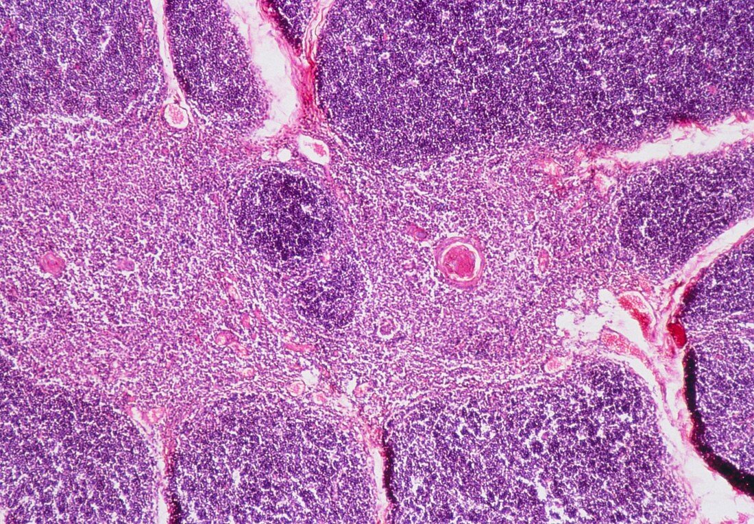
| Light micrograph of normal human thymus. The thymus is divided into lobules separated by septa of connective tissue (white spaces) which may contain blood vessels (as at right,stained red and very dark purple). Each lobule has a cortex (granular masses,densley packed,picture periphery) of lymphocytes,and a medulla (centre) of lymphocytes and stellate epitherial cells. The circular pink structures within the medulla (one prominent) are concentrically arranged epitherial cells of uncertain function. Magnificaton: x20 at 35mm size | |
| Licence : | Droits gérés |
| Crédit: | Science Photo Library / Michler, Astrid & Hans-Frieder |
| Taille de l’image : | 5079 px × 3530 px |
| Model Release : | Non requis |
| Property Release : | Non requis |
| Restrictions : | - |
Prix pour cette image À partir de 45 €
Produit vendu
(Calendrier, Carte postale, Carte de vœux, Impression sur textile, Packaging etc)
À partir de 45 €
Usage commercial
(Affichage, Annonce presse, Annonce TV, Carte, Digital - hors rés. sociaux, Digital - rés. sociaux etc)
À partir de 45 €
Éditorial
(Digital, Journal, Livre, Livre pratique, Magazine, Télévision etc)
À partir de 60 €
Usage non-commercial
(Digital - hors rés. sociaux, Digital - rés. sociaux etc)
À partir de 120 €